Download

1 / 40
420 likes | 1.02k Views
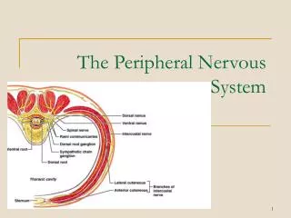
The Peripheral Nervous System. PART 3. Peripheral Nervous System. The Peripheral Nervous System. Nervous structures outside the brain and spinal cord Nerves allow the CNS to receive information and take action Functional components of the PNS

E N D
The PeripheralNervous System PART 3
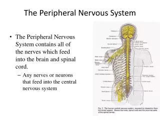
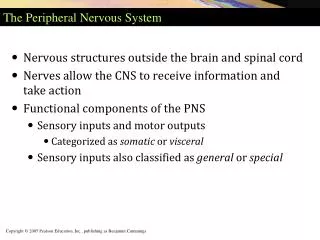
The Peripheral Nervous System • Nervous structures outside the brain and spinal cord • Nerves allow the CNS to receive information and take action • Functional components of the PNS • Sensory inputs and motor outputs categorized as somatic or visceral • Sensory inputs also classified as general or special
Sensory Input and Motor Output • Sensory (afferent) signals picked up by sensor receptors, carried by nerve fibers of PNS to the CNS • Motor (efferent) signals are carried away from the CNS, innervate muscles and glands • Divided according to region they serve • Somatic body region • Visceral body region • Results in four main subdivisions • Somatic sensory • Visceral sensory • Somatic motor • Visceral motor
PNS Afferent Division • Afferent (sensory) division – transmits impulses from receptors to the CNS. • Somatic afferent fibers – carry impulses from skin, skeletal muscles, and joints • Visceral afferent fibers – transmit impulses from visceral organs
PNS Efferent Division • Motor (efferent) division – transmits impulses from the CNS to effector organs. Two subdivisions: • Somatic nervous system – provides conscious control of skeletal muscles • Autonomic nervous system – regulates smooth muscle, cardiac muscle, and glands
Types of Sensory and Motor Information Figure 12.3
Sensory • General somatic senses – include touch, pain, vibration, pressure, temperature • Proprioceptive senses – detect stretch in tendons and muscle provide information on body position, orientation and movement of body in space • Special Senses - hearing, balance, vision, olfaction (smell), gustation (taste)
Motor • General somatic motor • Signals contraction of skeletal muscles • Under our voluntary control • Visceral motor • Makes up autonomic nervous system (ANS) • Regulates the contraction of smooth and cardiac muscle, controls function of visceral organs • ANS has two divisions • Parasympathetic • Sympathetic
Divisions of the ANS • Sympathetic - “fight or flight” • Catabolic (expend energy) • Mass activation prepares for intense activity. • Heart rate (HR) increases. • Bronchioles dilate. • Blood [glucose] increases. • Parasympathetic - “feed & breed”, “rest & digest” • Maintain homeostasis • Normally not activated as a whole, stimulation of separate parasympathetic nerves. • Relaxing effects: • Decreases HR. • Dilates visceral blood vessels. • Increases digestive activity. • Dual innervation of many organs — having a brake and an accelerator provides more control
Sympathetic Division Organization • Preganglionic neurons in segments T1 to L2 • Ganglia near the vertebral column • Sympathetic ganglia • Paired sympathetic chain ganglia • Unpaired collateral ganglia • Preganglionic fibers to adrenal medullae • Epinephrine (adrenalin)into blood stream
The Autonomic Nervous System • Effects of Sympathetic Activation • Generalized response in crises • Increased alertness/energy • Increased cardiovascular activity • Increased respiratory activity • Increased muscle tone
Parasympathetic Division Organization • Preganglionic neurons in brain stem and sacral spinal segment • Ganglionic neurons (peripheral ganglia) in or near target organ • Sacral fibers form pelvic nerves
The Autonomic Nervous System • Effects of Parasympathetic Activation • Relaxation • Food processing • Energy absorption • Brief effects at specific sites
Basic Structural Components of the PNS • Sensory receptors – pick up stimuli from inside or outside the body • Motor endings – axon terminals of motor neurons innervate effectors (muscle fibers and glands) • Nerves and ganglia • Nerves – bundles of peripheral axons • Ganglia – clusters of peripheral neuronal cell bodies
Nerves • Nerves – cablelike organs in the PNS • Consists of numerous axons wrapped in connective tissue • Endoneurium – layer of delicate connective tissue surrounding the axon • Perineurium – connective tissue wrapping surrounding a nerve fascicle • Nerve fascicles – groups of axons bound into bundles • Epineurium – whole nerve is surrounded by tough fibrous sheath • Axon is surrounded by Schwann cells
Cranial Nerves • Attach to the brain and pass through foramina of the skull • Numbered from I–XII • Cranial nerves I and II attach to the forebrain • All others attach to the brain stem • Primarily serve head and neck structures • The vagus nerve (X) extends into the abdomen
The 12 Pairs of Cranial Nerves Figure 14.5
Olfactory Nerves (I) • Sensory nerves of smell olfactory nerve (I) Olfactory bulb Olfactory tract Optic nerve (II) Optic chiasma Optic tract Oculomotor nerve (III) Trochlear nerve (IV) Trigeminal nerve (V) Abducens nerve (VI) Cerebellum Medulla Table 14.3 (1 of 12)
Optic Nerve (II) • Sensory nerve of vision Filaments of olfactory nerve (I) Olfactory bulb Olfactory tract Optic nerve (II) Optic chiasma Optic tract Oculomotor nerve (III) Trochlear nerve (IV) Trigeminal nerve (V) Abducens nerve (VI) Cerebellum Medulla Table 14.3 (2 of 12)
Oculomotor Nerve (III) • Innervates four of the extrinsic eye muscles Filaments of olfactory nerve (I) Olfactory bulb Olfactory tract Optic nerve (II) Optic chiasma Optic tract Oculomotor nerve (III) Trochlear nerve (IV) Trigeminal nerve (V) Abducens nerve (VI) Cerebellum Medulla Table 14.3 (3 of 12)
Trochlear Nerve (IV) • Innervates the superior oblique muscle (an extrinsic eye muscle) Filaments of olfactory nerve (I) Olfactory bulb Olfactory tract Optic nerve (II) Optic chiasma Optic tract Oculomotor nerve (III) Trochlear nerve (IV) Trigeminal nerve (V) Abducens nerve (VI) Cerebellum Medulla Table 14.3 (4 of 12)
Trigeminal Nerve (V) • Provides sensory innervation to the face • Motor innervation to chewing muscles Filaments of olfactory nerve (I) Olfactory bulb Olfactory tract Optic nerve (II) Optic chiasma Optic tract Oculomotor nerve (III) Trochlear nerve (IV) Trigeminal nerve (V) Abducens nerve (VI) Cerebellum Medulla
Abducens Nerve (VI) • Abducts the eyeball – innervates lateral rectus muscle Filaments of olfactory nerve (I) Olfactory bulb Olfactory tract Optic nerve (II) Optic chiasma Optic tract Oculomotor nerve (III) Trochlear nerve (IV) Trigeminal nerve (V) Abducens nerve (VI) Cerebellum Medulla Table 14.3 (6 of 12)
Facial Nerve (VII) • Innervates muscles of facial expression Facial nerve (VII) Vestibulocochlear nerve (VIII) Glossopharyngeal nerve (IX) Vagus nerve (X) Accessory nerve (XI) Hypoglossal nerve (XII) Table 14.3 (7 of 12)
Vestibulocochlear Nerve (VIII) • Sensory nerve of hearing and balance Facial nerve (VII) Vestibulocochlear nerve (VIII) Glossopharyngeal nerve (IX) Vagus nerve (X) Accessory nerve (XI) Hypoglossal nerve (XII) Table 14.3 (8 of 12)
Glossopharyngeal Nerve (IX) • Innervates structures of the tongue and pharynx Facial nerve (VII) Vestibulocochlear nerve (VIII) Glossopharyngeal nerve (IX) Vagus nerve (X) Accessory nerve (XI) Hypoglossal nerve (XII) Table 14.3 (9 of 12)
Vagus Nerve (X) • A mixed sensory and motor nerve - “Wanders” into thorax and abdomen Facial nerve (VII) Vestibulocochlear nerve (VIII) Glossopharyngeal nerve (IX) Vagus nerve (X) Accessory nerve (XI) Hypoglossal nerve (XII) Table 14.3 (10 of 12)
Accessory Nerve (XI) • An accessory part of the vagus nerve -innervates trapezius muscle Facial nerve (VII) Vestibulocochlear nerve (VIII) Glossopharyngeal nerve (IX) Vagus nerve (X) Accessory nerve (XI) Hypoglossal nerve (XII) Table 14.3 (11 of 12)
Facial nerve (VII) Vestibulocochlear nerve (VIII) Glossopharyngeal nerve (IX) Vagus nerve (X) Accessory nerve (XI) Hypoglossal nerve (XII) Hypoglossal Nerve (XII) • Runs inferior to the tongue - innervates the tongue muscles Table 14.3 (12 of 12)
Spinal Nerves • 31 pairs – contain thousands of nerve fibers • Connect to the spinal cord • Named for point of issue from the spinal cord • 8 pairs of cervical nerves (C1–C8) • 12 pairs of thoracic nerves (T1–T12) • 5 pairs of lumbar nerves (L1–L5) • 5 pairs of sacral nerves (S1–S5) • 1 pair of coccygeal nerves (Co1)
Spinal Nerves • Connect to the spinal cord by the dorsal root and ventral root • Dorsal root – contains sensory fibers • Dorsal root ganglion – of afferent cell bodies • Ventral root – contains motor fibers arising from anterior gray column • Branch into dorsal ramus and ventral ramus both contain sensory and motor fibers • Rami communicantes connect to the base of the ventral ramus and lead to the sympathetic chain ganglia
White matter Ventral root Gray matter Dorsal root Dorsal root ganglion Dorsal and ventral rootlets of spinal nerve Dorsal ramus of spinal nerve Spinal Nerves Ventral ramus of spinal nerve Spinal nerve Rami communicantes Sympathetic trunk (chain) ganglion (a)
Innervation of the Back • Dorsal rami • Innervate back muscles • Follow a neat, segmented pattern • Innervate a horizontal strip of muscle and skin • In line with emergence point from the vertebral column
Innervation of the Thoracic region • Ventral rami arranged in simple, segmented pattern • Intercostal nerves – supply intercostal muscles, skin, and abdominal wall • Each gives off lateral and anterior cutaneous branches
Introduction to Nerve Plexuses • Nerve plexus – a network of nerves • Ventral rami (except T2 – T12) • Branch and join with one another • Form nerve plexuses • Cervical • Brachial • Lumbar • Sacral • Primarily serve the limbs • Fibers from ventral rami crisscross
The Cervical Plexus • Buried deep in the neck under the sternocleidomastoid muscle • Formed by ventral rami of first four cervical nerves (C 1 – 4) • Most are cutaneous nerves • Some innervate muscles of the anterior neck
Brachial Plexus • Brachial plexus lies in the neck and axilla • Formed by ventral rami of C5 – C8 give rise to cords • Cords give rise to main nerves of the upper limb Figure 14.9d
Lumbar Plexus • Arises from L1– L4 • Smaller branches innervate the posterior abdominal wall and psoas muscle • Main branches innervate the anterior thigh
The Sacral Plexus • Arises from spinal nerves L4–S4 • Often considered with the lumbar plexus referred to as the lumbosacral plexus • Sciatic nerve – the largest nerve of the sacral plexus is actually two nerves in one sheath • Tibial nerve – innervates most of the posterior lower limb • Common fibular (peroneal) nerve – innervates muscles of the anterolateral leg