The Peripheral Nervous System
860 likes | 2.24k Views
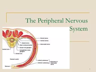
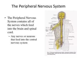
The Peripheral Nervous System. The Peripheral Nervous System contains all of the nerves which feed into the brain and spinal cord. Any nerves or neurons that feed into the central nervous system. The Peripheral Nervous System. Somatic Nervous System

The Peripheral Nervous System
E N D
Presentation Transcript
The Peripheral Nervous System • The Peripheral Nervous System contains all of the nerves which feed into the brain and spinal cord. • Any nerves or neurons that feed into the central nervous system
The Peripheral Nervous System • Somatic Nervous System • The division of the peripheral nervous system that controls the body’s skeletal muscles • Autonomic Nervous System • The part of the peripheral nervous system that controls the glands and the muscles of the internal organs (such as the heart) • Sympathetic Nervous System • The division of the autonomic nervous system that arouses the body, mobilizing its energy in stressful situations • Parasympathetic Nervous System • The division of the autonomic nervous system that calms the body, conserving its energy
Reflexes • Our automatic response to stimuli arereflexes. • A simple spinal reflex pathway is composed of a single sensory neuron and a single motor neuron, connected through the spine with an inter neuron. • This type of response does not involve the brain, and is often why we feel our body move before we feel the stimuli • A warm, headless body could demonstrate a reflex like that produced when hitting the patellar tendon with a hammer.
A. Afferent neuron B. Efferent neuron C. Interneuron
Nervous System Peripheral Nervous System (PNS) Central Nervous System (CNS) Autonomic System Somatic System Sympathetic (Arousing) Parasympathetic (Calming) Divisions of the Nervous System
The Endocrine System The endocrine system is the body’s chemical messenger system, that relies on hormones. It involves the endocrine glands: pituitary, thyroid, parathyroid, adrenals, pancreas, ovaries, and testes. Hormones are chemical messengers used by the endocrine system. Many hormones are also neurotransmitters.
Working with Other Systems • Under normal (unaroused) conditions, the endocrine system works in parallel with the parasympathetic nervous system to sustain our basic body processes. • In crisis, the endocrine system shifts into a new mode to support the sympathetic nervous system….it releases epinephrine (adrenalin) • Triggers the “fight or flight” response
The Master Gland • While the body has a many glands which are important, the most important glad is the pituitary gland. • Controls all of the responses of the endocrine system • The pituitary gland is no larger than a pea, and is located at the base of the brain.
The Brain • To get a feel for how complex our brains are think about this: • You could join two eight-studded Lego bricks 24 ways, and six bricks nearly 103 million ways. With some 40 billion neurons, each having roughly 10,000 contacts with other neurons, we end up with around 400 trillion synapses. • A grain of sand size speck of your brain contains 100,000 neurons and one billion synapses.
Neurons in the brain connect with one another to form networks Inputs Outputs The brain learns by modifying certain connections in response to feedback The Brain • Neurons cluster into work groups called neural networks. • To understand why this happens, think about why cities exist and how they work. • Neurons work with those close by to ensure short, fast connections.
The Brain • For creatures with more complex brains, there are three levels. Creatures with complex brains all share a similar stalk, the brain stem. • The brain stem is the part of the brain with the longest ancestry. • On top of the brain stem, in more evolved creatures, are the limbic system and the cerebral cortex.
The Brain Stem The brain stem is made up of four regions: the medulla, the pons, the reticular formation and the thalamus.
The Medulla The medulla is the bulge low in the brain stem. It regulates basic body functions including breathing, blood pressure and heart rate. The medulla operates on autopilot without our conscious awareness.
The Pons • The pons is an even larger bulge that sits just above the medulla. • The pons helps relay signals to the cerebellum, that deal with sleep, respiration, swallowing, bladder control, hearing, equilibrium, taste, eye movement, facial expressions, facial sensation, and posture. • Pons is Latin for bridge; a fitting name since it acts as a “bridge” which connect the brain stem to the cerebellum.
The Reticular Formation • The reticular formation is a pencil shaped bundle of nerve cells that forms the brain stem’s core. • One of the reticular formation’s jobs is to keep the brain awake and alert. • Also is responsible for monitoring incoming sensory messages.
The Thalamus The thalamus is at the very top of the brain stem and lays near the center of the brain. The thalamus is like the central processing chip of a computer and directs all incoming and outgoing sensory and motor traffic.
The Cerebellum Sometimes called the “little brain,” the cerebellum sits at the back of the brain stem and looks like a miniature version of our brain. It coordinates with the brain stem and higher parts of the brain to control complex movements we perform without consciously thinking about-walking, dancing, or drinking from a cup.
The Cerebellum Acting with the brainstem, the cerebellum controls the most basic functions of movement and life itself. Most of the work it does is automatic, and occurs outside out consciousness.
Limbic System The limbic system is the middle layer of brain that wraps around the thalamus. Together, the limbic system and the thalamus give humans/mammals the capability for emotions and memory
Limbic System • The layers of the limbic system not only processes memories and regulate emotions, it is also involved in feelings of pleasure, pain, fear and rage. • Expands on the more basic functions of the brain stem.
Hippocampus One of the two important parts of the limbic system is the hippocampus. Technically there are two hippocampi and their job is to connect your present with your past memories.
Amygdala The second part of the limbic system that is important is the amygdala. Like the hippocampi, the amygdalas’ job relates to memory and emotion. It also seems to play the largest role in dealing with feelings of pleasure.
Hypothalamus • A third part of the limbic system is the hypothalamus. It’s function is to analyze the blood flow in your body. • Specifically regulates body temperature, fluid levels and nutrients. • When it detects an imbalance, it tells the body how to respond. • Feeling thirsty or hungry.
Cerebral Cortex Major Lobes of the Brain When you look at a human brain, the majority of what you see is the cerebral cortex.
Frontal and Parietal Lobes • Frontal Lobes: Portion of the cerebral cortex just behind the forehead. • Involves the motor cortex. • Involved in making plans and judgment. • Parietal Lobes: Portion of the cerebral cortex at the top of the head. • Used for general processing, especially mathematical reasoning.
Temporal and Occipital Lobes • Temporal Lobes: The temporal lobe is involved in auditory processing. • It is also heavily involved in semantics both in speech and vision. • The temporal lobe contains the hippocampus and is therefore involved in memory formation as well. • Occipital Lobes:Portion of the cerebral cortex just at the back the brain • Responsible for visual functions
Broca and Wernicke • Broca’s Area: Located in the left frontal lobe. • Is involved with expressive language. • Damage to this area results in difficulty with spoken language. • Area directs muscle movements important to speech production. • Wernicke’s Area: Located in the temporal lobe. • Controls receptive language (understands what someone else says.)
Aphasia • Damage to any one of several cortical areas can cause aphasia, or an impaired use of language. • When you read words aloud, the words (1) register in the visual area, (2) are relayed to a second area, the angular gyrus, which transforms them into an auditory code that is (3) received and understood in Werneicke’s area and (4) sent to Broca’s area, which (5) controls the motor cortex as it creates the pronounced word. Depending on which link in the chain is damaged, a different form of aphasia occurs.
Damage to Broca’s Area • Another example of this could be Foreign Accent Syndrome, or FAS. • When a person experiences brain damage in Broca’s area, the result is often times expressed in difficulty with speech. • Common in stroke patients
Motor Cortex • Motor Cortex: An area of the brain at the back of the frontal lobe. • In charge of the movement of your body parts. • The motor cortex on the right side of your brain controls the movement of the left side of your body, and vice versa. • The more intricate the movement for 1 body part, the bigger the section on the motor cortex. • SomatosensoryCortex: The are just behind the motor cortex where your body registers and processes sensations. • Association Areas: areas that associate various sensory inputs with stored memory.
Primary motor cortex (M1) Hip Trunk The Motor Cortex Arm Hand Foot Face Tongue Larynx
Cerebral Dominance • Right Hemisphere • Regulation of negative emotions. • Response to simple commands. • Memory for shapes and music. • Interpreting spatial relationships and visual images. • Recognition of faces. • Left Hemisphere • Regulation of positive emotions. • Control of muscles used in speech. • Control of sequence of movements. • Spontaneous speaking and writing. • Memory for words and numbers. • Understanding speech and writing. While both sides of the brain rely on the other half, each hemisphere of the cerebral cortex has specific functions.
Cerebral Dominance Keeping in mind that the left side of the brain controls the right side of the body, and vise-versa, we must understand that an injury to the left side of the brain will show bodily symptoms on the right side. We also must keep in mind that while each side of the brain may be responsible for certain actions and abilities, the two areas work cooperatively on most tasks.
Hemispheric Differences • One common misconception is that people can be “right brained” or “left brained.” • This is another example of pseudo-psychology. In reality we use the both sides of our brain, and the communication between the two halves is important. • “Right Brain”/”Left Brain” test
The Splint Brain Procedure The Split Brain Experiment In the recent past, patients who had severe cases of epilepsy would sometimes be treated with a procedure they called the “split brain.” In this procedure they would literally cut the brain in two by cutting the corpus collosum.
The Split Brain Procedure The Split Brain Procedure For these patients, life changed very little on the service, with the exception of far fewer seizures. Put under certain circumstances, however, the side effects were very clear.
The Endocrine System • Endocrine System • The body’s “slow” chemical communication system • A set of glands that secrete hormones into the bloodstream
EEG (Electroencephalography) • Technique: Multiple electrodes are pasted to outside of head • What it shows: A single line that charts the summated electrical fields resulting from the activity of billions of neurons
EEG (Electroencephalogram) • Advantages • Detects very rapid changes in electrical activity, allowing analysis of stages of cognitive activity • Disadvantages • Provides poor spatial resolution of source of electrical activity
PET(Positron Emission Tomography)SPECT(Single Photon Emission Computed Tomography) • Technique: Positrons and photons are emissions from radioactive substances • What it shows: An image of the amount and localization of any molecules that can be injected in radioactive form, such as neurotransmitters, drugs, tracers for blood flow or glucose use (which indicates specific changes in neuronal activity) PET Scan
PET(Positron Emission Tomography)SPECT(Single Photon Emission Computed Tomography) • Advantages • Allows functional and biochemical studies • Provides visual image corresponding to anatomy • Disadvantages • Requires exposure to low levels of radioactivity • Provides spatial resolution better than that of EEG, but poorer than that of MRI • Cannot follow rapid changes (faster than 30 seconds)