Download
1 / 80
900 likes | 1.33k Views
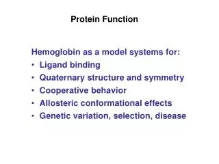
Protein Function and Evolution. Role of Globins in O 2 Transport and Storage. Red Blood Cells (Erythrocytes). Myoglobin (Mb) and Hemoglobin (Hb). Structures of Porphyrins. Ferrous iron, Fe 2+ , in heme binds O 2 .

E N D
Structures of Porphyrins Ferrous iron, Fe2+, in heme binds O2. Heme also has high affinity for other molecules, such as carbon monoxide. This is why CO is toxic.
Iron in Oxyhemoglobin Heme bound to protein as prosthetic group (tightly bound co-factor) protects heme iron from oxidation (from Fe2+ to Fe3+ oxidation state), lowers heme’s extremely high affinity for CO, and allows for regulation of O2 binding affinity in hemoglobin. His93 (F8) = proximal His His64 (E7) = distal His
Mb + O2 <-> MbO2 K (association constant) = [MbO2]/([Mb][O2]) (theta; fractional occupancy) = sites occupied/total available sites = [MbO2]/([Mb]+[MbO2]) = K[Mb][O2]/([Mb]+ K[Mb][O2]) = K[O2]/(1+ K[O2]) = [O2]/(1/K + [O2]) 1/K = Kd (dissociation constant) = P50 (O2 partial pressure for half-maximal saturation), so = PO2/(P50 + PO2) Equations for Myoglobin Binding O2
Dynamics of 02 Release by Myoglobin Kd = 1/K = ([Mb][O2])/[MbO2] = koff/kon von(on rate) = kon[O2][Mb] voff(off rate)= koff[MbO2]
T (“tense”) conformational state: low-affinity ligand-binding state of protein. R (“relaxed”) conformational state: high-affinity binding binding state of protein. Homotropic allosteric interaction: effector and ligand regulated by the effector are the same molecule (e.g., O2 binding affects subsequent O2 binding). Heterotropic allosteric interaction: effector and ligand are different molecules (e.g., H+ or BPG binding affects O2 binding). Positive allosteric interaction: effector binding increases affinity for ligand. Negative allosteric interaction: effector binding decreases affinity for ligand. Cooperative Binding and Allostery allostery = “other site”
Evaluating Cooperativity Fractional O2 occupancy: = PO2n/(P50n + PO2n) Rearrange and take logarithms for Hill plot: log(/(1-)) = nlogP02 - nlogP50 (assumes n = # of O2 binding sites & all O2 bind simultaneously) In fact, experimentally determined “n” value is Hill coefficient (nH). nH n except for hypothetical, wholly cooperative process where all ligand molecules would bind simultaneously. nH < n for all real systems. Hill coefficient very useful in describing cooperativity, since: nH = 1: non-cooperative process nH > 1: positive cooperativity nH < 1: negative cooperativity Archibald Hill (1910)
Hill Plots for O2 Binding for Mb and Hb For Mb: nH = 1 This indicates non-cooperative process. For Hb: nH (max slope) = 3.0-3.5 This indicates positive cooperativity (since nH > 1) with at least 4 binding sites for O2 (since n always > nH). • Intercepts with broken black line at the 0 value for log(/(1-)) indicate P50 and so O2 binding affinity (lower P50 =higher affinity) • Hb high-affinity 02-binding logP50 = upper asymptote intercept • Hb low-affinity 02-binding logP50 = lower asymptote intercept
Bound ligand/free ligand vs. bound ligand X-axis intercept indicates maximum amount of ligand bound (Bmax) or total number of ligand binding sites (n), e.g., 1 for Mb, 4 for Hb. Slope = -K = -1/Kd (or -1/P50) Scatchard Plots Shape of curve gives indication of whether there is cooperativity (positive or negative) or not. Note: Scatchard plots and other data plotting methods in biochemistry are used a great deal for visual/graphical representation even today. Biochemical parameters used to be determined by manual plotting but now computers are used, since regression analysis on a computer is much more accurate for determining n, Kd, etc.
Some Major Interactions in deoxyHb that Are Disrupted in T -> R Transition to oxyHb
Some Major Interactions in deoxyHb that Are Disrupted in T -> R Transition to oxyHb
Changes at a1-b2 and a2-b1 Interface in T -> R Transition in Hb
Change in Hb 4o Structure with O2 Binding • Some major changes: • Rotation of a1b1 relative to a2b2. • Change in size of central cavity. • Shift of C-termini and FG corners of b chains relative to C helices of a chains • C-termini of b chains interact with C helices of a chains in T (deoxy) state. These interactions are broken in transition to R (oxy) state.
Mechanism of T -> R Transition: Iron Pulled into Heme Plane when O2 Binds His F8 (proximal His) also dragged along in T -> R transition, pulling F helix and shifting subunits relative to one another, increasing O2 affinity of binding sites on other subunits. Perutz Model (1970)
Effect of Replacing Proximal His in Hb with Gly and Adding Imidazole Normal Hb: Cooperativity Replacement: No cooperativity
H+ (“The Bohr effect,” Christian Bohr, 1904) 2,3-bisphosphoglycerate (BPG) Carbon dioxide (transported in blood as bicarbonate and carbamates): Bicarbonate formation: CO2 + H20 <-> HCO3- + H+ Carbamate formation: Hb-NH3+ +HCO3- <=> Hb-NH-COO- + H+ + H2O CO2 lowers O2 binding affinity through H+ released (contributing to Bohr effect) and formation of carbamate at N-termini of Hb b subunits, stabilizing T state interactions between a and b chains. Negative Allosteric Effectors of O2 Binding in Hb: Stabilizers of T State of Hb
Bohr Effect on Hb: Protonation of Certain Groups on Hb Decreases Affinity for O2 Protonation of a number of groups favors T state. For instance: Protonation of His146 (HC3) on b chain allows for formation of T (deoxy) state salt bridge with Asp94.
Networks of Ion Pairs and Hydrogen Bonds in DeoxyHb All of these interactions are broken in T -> R transition. (White + signs: groups protonated in Bohr effect, stabilizing deoxyHb T state.)
2,3-Bisphosphoglycerate (BPG) In mammals In birds
Release of CO2 in lungs (or gills in fish). Oxygenation of Hb in lungs. Role of Globins in O2 Transport and Storage CO2 carried in veins as bicarbonate. Also, deoxyHb carries CO2 as carbamates. OxyHb carries O2 in arteries. CO2, H+ and BPG decrease Hb’s affinity for O2 and so favor release of O2 in tissues. [CO2] (and [H+]) high in tissues as a result of respiration.
Two Models of Allostery • Koshland, Nemethy, Filmer (KNF) Model (1966): Sequential or Induced Fit Model • Ligand binding at one site causes protein conformational change (induced fit), shifting binding affinity in adjacent subunits only, so complete T -> R transition is a sequential process. • Can account for both positive and negative cooperativity. • Monod, Wyman, Changeux (MWC) Model (1965): • Concerted or Symmetry Model • Equilibrium between T and R states. • Transition is a concerted process, affecting all subunits simultaneously in the same way. • In absence of ligand, equilibrium favors T state. • Ligand binding shifts equilibrium toward R state. • Only models positive cooperativity.
Two Models of Allostery Koshland, Nemethy, Filmer (KNF) Model (1966): Sequential or Induced Fit Model Monod, Wyman, Changeux (MWC) Model (1965): Concerted or Symmetry Model
Recent Model for Cooperative Transition of Hb If both a1b1 and a2b2 each contain at least one O2 bound, T -> R transition occurs.