PROTEIN STRUCTURE AND FUNCTION
PROTEIN STRUCTURE AND FUNCTION. Proteins Are Where It’s At. Proteomics Gene regulation Drug Discovery Understanding evolution Etc. Proteins are Where It’s Been. Enzymes ß-galactosidase Antibodies Anti-Hepatitis B Hormones Human Growth Hormone Estrogen Testosterone.

PROTEIN STRUCTURE AND FUNCTION
E N D
Presentation Transcript
Proteins Are Where It’s At • Proteomics • Gene regulation • Drug Discovery • Understanding evolution • Etc.
Proteins are Where It’s Been • Enzymes • ß-galactosidase • Antibodies • Anti-Hepatitis B • Hormones • Human Growth Hormone • Estrogen • Testosterone
Proteins are Where It’s Been • Structural proteins • Collagen • Transportation • Haemoglobin
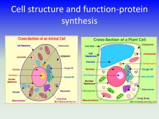
Proteins Are Us • In cells, when something needs to be done, it is a protein that does it. • The human body contains over 30,000 different types of protein • Other organisms have many of the same proteins as well as different ones • Enzymes are the biggest class • 3,000 enzymes in average mammalian cell • ß-galactosidase is an enzyme
Transcription Factors Control expression of genes Hormones Control body function Antibodies Fight infection Enzymes Speed up chemical reactions Carrier molecules Haemoglobin -carries oxygen in the blood Structural Collagen Found in bone and skin Keratin ■ Makes hair and nails Fibrin Helps clot blood Elastin Major part of ligaments Classes of Proteins
Proteins AreDiverse In Structure • Proteins can do many things because they are structurally diverse • Differ in many properties: • Size • Shape • Charge distribution • Hydrophobicity • Solubility properties
Variability ComesFrom Amino Acids • Are polymers composed of 20 different amino acid building blocks • As letters can be arranged in many ways, so too can amino acids • Number, type and arrangement of amino acids determines structure and function • Insulin has about 50 AA • Most are >> bigger - from 100s to 1000s • Allows for great diversity
Amino Acids • All amino acids have a carboxyl group and an amino group • A different R group is attached to each amino acid
R groups make each amino acid different • Some are: • Polar • Nonpolar • Charged • Acids • Bases
Folding • DNA always has same structure • But proteins fold into many different shapes • Folding depends ultimately on amino acid composition • Structure of proteins determines function • Structure allows proteins to • BIND to other molecules • RECOGNIZE other molecules
Shape Is Critical • Change in one amino acid can change the structure of the protein with a large effect on function • Sickle cell anemia
Sickle Cell Anemia • Single DNA base pair is mutated • Therefore one amino acid is altered • Glutamic acid is switched to valine • Glutamic acid is negatively charged, valine is neutral • Changes how hemoglobin packs in cells • Alters shape of red blood cells when oxygen is low. Image fromMedline Plus
Levels of ProteinStructure • Protein structure is complex and important, so it is classified into: • 1° - Primary • 2°- Secondary • 3° -Tertiary • 4°- Quaternary
Primary Structure • Linear sequence of amino acids • Peptide bond (covalent bond) holds it together • Beads on a string
Peptide Bonds R O H R l ll l l NH2 –C – C – 0 –H + H –N – C—COOH l l H H R O H R l ll l l NH2 -- C –C – N – C – COOH + H2O l l H H
Secondary Structure • Regular repeating patterns of twists or kinks of the amino acid chain • Examples • Alpha helix • Beta pleated sheet
Secondary Structure • Hydrogen bonds hold structure together • Weak, noncovalent, molecular interactions • Hydrogen atom is bonded to an electronegative atom (like F, O, N) that is also partially bonded to another atom (usually also F, O, N)
Figure from National Human Genome Research Institute, by artist Darryl Leja. Used with permission.
TertiaryStructure 3° • 3-D Globular Configuration formed by bending and twisting of the polypeptide chain • Stabilized by: • Hydrogen bonds • Electrostatic interactions • (Positive and negative) • Hydrophobic interactions • Sometimes covalent bonds • Disulfide bonds
QuaternaryStructure 4° • Two or more polypeptide chains associate with each other
ß-Galactosidase • Link to Protein Data Bank for picture of molecular image of ß-galactosidase • www.pdb.org
Higher OrderStructure • Higher order (secondary, tertiary, quaternary) structure is relatively “weak” • In nature, “weakness” of noncovalent interactions is important • Flexibility • Enzymes change shape when binding to their substrates • Necessary for proper function
How Proteins Lose Normal Structure And Function • Primary structure hard to disrupt; covalent bonds are strong
How Proteins Lose Normal Structure And Function • Can be broken apart by enzymes (proteases) that digest the covalent peptide bonds • Called proteolysis • Occurs naturally in digestion • Can be a problem in the lab; proteases can destroy protein of interest • Use cold to avoid proteolysis
How Proteins Lose Their Structure And Function • Sulfur groups on cysteines may undergo oxidation to form disulfide bonds that are not normally present • Proteins can aggregate leading to precipitation • Proteins can adsorb (stick to) surfaces
Higher OrderStructure In Lab • Loss of higher order structure is denaturation • Denaturation occurs fairly easily • Affected by changes in pH • Ionic strength • Temperature • May or may not be reversible
ManipulatingHigher Order Structure • Often manipulated in lab • Destroy folding when we do PAGE • Use buffers to maintain the structure • Use cold temperatures • Add reducing agents to prevent unwanted disulfide bonds in the lab --DTT or -ME
Analyzing Protein Structure • X-Ray Crystallography • Like a CAT scan in medicine • X-ray taken at multiple angles and computer uses the data to calculate a 3D image • Nuclear Magnetic Resonance • Like an MRI in medicine
X-rayCrystallography • Isolate and purify protein • Form a crystal of the protein • Molecules of the protein are arranged in an orderly lattice • Dissolve protein in solvent • Precipitate into a crystal • X-Ray the crystal
X-rayCrystallography • Analyze diffraction pattern using software • Make electron density map • Process used to take years • Different versions of crystal for comparison • Each with different heavy metal in lattice to provide reference point • New X-ray sources - synchrotrons have reduced data collection time to few days
X-rayCrystallography • Synchotron - Argonne National Lab • Still takes weeks to go from gene sequence to 3-D structure • (mrsec website, nanotechnology at UW-Madison)
StructuralGenomics • Goal to solve thousands of structures a year • Large scale automation required • Syrrx - structural genomics company • Robot places a drop of protein into 480 wells • 11,000 crystallization experiments in 24 hours • New robot - 130,000 a day
Nuclear MagneticResonance • Similar to MRI - Magnetic Resonance imaging in medicine • No need for crystals, proteins in solution • Works for relatively simple proteins