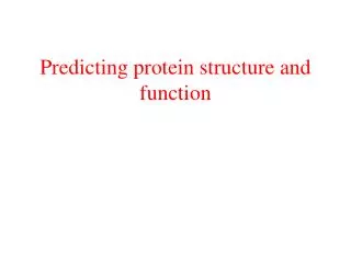
Protein Structure and Function
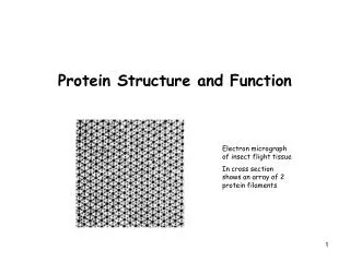
Protein Structure and Function. Electron micrograph of insect flight tissue In cross section shows an array of 2 protein filaments. Structure and Flexibility indicates Function. DNA polymerase III – DNA complex (Replication). Conformational change of lactoferrin upon binding of Fe.

Protein Structure and Function
E N D
Presentation Transcript
Protein Structure and Function Electron micrograph of insect flight tissue In cross section shows an array of 2 protein filaments
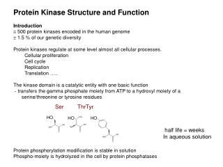
Structure and Flexibility indicates Function DNA polymerase III – DNA complex (Replication) Conformational change of lactoferrin upon binding of Fe
Proteins are Polypeptides Direction of a Protein
Cys can cross-link between 2 polypeptide chains -> Disulfide bridge Covalent cross-link on 3° structure level
α-helical coiled coil proteins: Form superhelix Found in myosin, tropomyosin (muscle), fibrin (blood clots), keratin (hair) The cytoskeleton is rich in filaments which are α-helical coiled coil proteins Examples of α-Helical Proteins:
Examples of α-Helical Proteins: Bacteriorhodopsin (Photoreceptor) Many membran proteins are α-helical
Examples of β-sheet Proteins: Fatty acid binding protein -> β barrels structure OmpX: E. coli porin Antibodies
Quaternary Structure: Polypeptide chains assemble into multisubunit structures Cell-surface receptor CD4 Cro protein phage λ Tetramer of hemoglobin Coat protein of rhinovirus
Protein Folding Folding is a highly cooperative process (all or none) Folding by stabilization of Intermediates
Protein Modifications GFP fluorescent: Rearrangement and oxidation of Ser-Tyr-Gly
Protein Trafficking Bovine cell stained with fluorescent dyes. Green -> ER Red -> Mitochondria
Secretory proteins are transported to ER shortly after synthesis started
Synthesis of secretory proteins and their cotranslational translocation across the ER membrane
Synthesis of secretory proteins and their cotranslational translocation across the ER membrane • What is needed for translocation: • Signal sequence (9-12 hydrophobic AA with some mainly pos. charged ones – in some prokaryotes sometimes longer, most of the times cleaved off by peptidases on the ER lumen side, sequence mainly at N-terminal) • Signal-Recognition-Particle (SRP) –recognizes signal sequence of ribosome complex (ribosome with mRNA), redirects ribosome complex to SRP receptor, puts synthesis of protein on hold • SRP receptor – binds the ribosome- SRP complex - driggers that ribosome complex is moved to translocon (GTP dependent) • Translocon is a protein channel, opens upon binding of ribosome complex, synthesis through channel
N-terminal signal sequence of secretary and membrane proteins
Post-translational Translocation Fairly common in yeast and occationally in higher eukaryotes.
Synthesis and insertion into the ER of membrane proteins Type I Type II
GPI-anchored Proteins • Glycosylphosphatidylinositol (GPI) • From yeast • In other organisms -> differs in • Acyl chain • Carbohydrate moiety Formation of GPI-anchored proteins in the ER membrane
Protein Modification • Membrane and soluble secretary proteins synthesized on the ER have 4 possible modifications before the reach final destination: • Glycosylation in ER and Golgi • Formation of S-S bonds in ER • Proper folding and assembly of multisubunits in ER • Proteolytic cleavage in ER, Golgi, and secretory vesicles
Protein Modification - Glycosylation O-linked glycosylkation: Attachment of sugars to OH of Ser and Thr Often contain only 1-4 sugar groups N-linked glycosylation: Attachment of sugars to amine N of Asn (Asn-X-Ser/Thr) Larger and more sugar groups -> more complex Glycosylation patters differ slightly between spieces !!! In Yeast: N-linked glycosylation are classified as core and mannan types. The core type contains 13-14 mannoses whereas the mannan-type structure consists of an inner core extended with an outer chain of up to 200-300 mannoses, a process known as hyperglycosylation. Precursor of N-linked sugars that are added to proteins in the ER
Processing of N-linked glycoproteins in the Golgi apparatus of mammalien cells Galactose addition + neuraminic acid linkage to galactose Gucosamine addition Mannose trimming
Pathways for formation of S-S bonds in Eukaryotes and Bacteria
Folding and assembly of Multimers Hemagglutinin trimer folding Binding of Chaperone BiP Closing S-S bond, N-linked glycosylation Membrane anchoring Assembly of trimer Another example for assembly of multimers -> immunoglobulins
Improperly Folded Protein Induce Expression of Chaperons Unfolded and incomplete folded protein in the ER -> releases chaperons (BiP) from Ire1 -> upon release of BiP Ire1 dimerizes (activation) -> Endonuclease activity in th cytosol -> splices Transcription factor Hac1 -> Hac1 protein returns into nucleus -> activates transcription of Chaperons -> Misfolded and unassembled proteins -> transported from the ER to the cytosol -> degradation
Modification of Proteins - Proteolytic Cleavage Proteolytic cleavage of proinsulin occurs in secretory vesicles (after Golgi)
Export of Bacterial Proteins Post-translational translocation across inner membrane of gram-negative bacteria
Injection of Protein by Pathogenic Bacteria (into Animal cells) Yersinia pestis: Causes Pest Virulence: Disables host macrophages -> by injecting a small set of proteins into macrophages Secretion mechanism for injecting bacterial proteins into Eukaryotic cells
Protein Transport between Organelles are done by Vesicles Assembly of protein coat drives vesicle formation and selection of cargo molecules
Assembly and Disassembly of Coat protein Interaction of cargo protein with vesicle N-terminus of Sar1 (membrane anchor) not shown
Model for Docking and Fusion of Transport vesicles with Target Membrane
Vesicle-mediated Protein Trafficking between ER and Golgi Backtransport mainly used for: -> recycling of membrane bilayer -> recycling of proteins (SNARE) -> missorted proteins Normal transport of secretory proteins
Involvement of the 3 major types of coat proteins in traffic and secretory pathways
Membrane Fusion directed by Hemagglutinin (HA) Influenza Virus: Glycoprotein on suface of virus After endocytosis (uptake of virus of the cell) viral envelop fuses with endosomal membrane Acidic pH necessary for conformational change in HA -> viral HA can insert into endosomal membrane