Download
1 / 10
150 likes | 445 Views
Cerebrospinal fluid. CSF. In adults, approximately 500 mL of cerebrospinal fluid (CSF) is produced each day (0.3-0.4 mL /min). The total adult volume varies from 90-150 mL. In neonates, the volume varies from 10-60 mL. Thus, the total CSF volume is replaced every 5-7 hours.

E N D
In adults, approximately 500 mL of cerebrospinal fluid (CSF) is produced each day (0.3-0.4 mL/min). • The total adult volume varies from 90-150 mL. • In neonates, the volume varies from 10-60 mL. • Thus, the total CSF volume is replaced every 5-7 hours
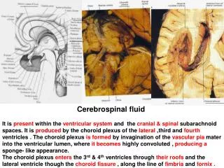
The CSF has several major functions: • (a) it provides physical support since the 1500 g brain weighs about 50 g when suspended in CSF; • (b) it confers a protective effect against sudden changes in acute venous and arterial blood pressure or impact pressure; • (c) it provides an excretory waste function since the brain has no lymphatic system; • (d) it is the pathway whereby hypothalamus releasing factors are transported to the cells; • (e) it maintains central nervous system ionic homeostasis.
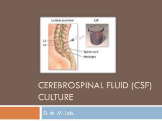
Specimen Collection • Cerebrospinal fluid may be obtained by lumbar puncture,or spinal tap . A spinal needle is inserted, usually between the 3rd and 4th lumbar vertebrae in the lower spine. Once the needle is properly positioned in the subarachnoid space (the space between the spinal cord and its covering, the meninges), pressures can be measured and fluid can be collected for testing.
The CSF specimen is usually divided into three serially collected sterile tubes: • tube 1 for chemistry and immunology studies; • tube 2 for microbiologic examination; and • tube 3 for cell count and differential.
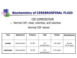
Normal CSF is crystal clear and colorless and has a viscosity similar to that of water. • Abnormal CSF may appear cloudy, frankly purulent, or pigment tinged. • Turbidity or cloudiness begins to appear with leukocyte (WBC) counts over 200 cells/μL or red cell (RBC) counts of 400/μL.
Appearance • If CSF is not crystal clear, a pathologic condition of the CNS should be suspected • Compare fluid to water • Fluid may be clear with as many as 400 RBCs/mm3 and 200 WBCs/mm3
Cells • WBC counts over 5 cells/mm3 should be taken to indicate the presence of pathologic condition • Polymorphonuclear leukocytes are never seen in normal adults • Neutrophilicpleocytosis is commonly associated with bacterial infections or early stages of viral infections, tuberculosis, meningitis, hematogenous meningitis, and chemical meningitis due to foreign bodies.
Glucose • Low CSF glucose concentration indicates increased glucose use in the brain and the spinal cord. • The normal range of CSF glucose is between 50 and 80 mg/dl • 60-70% of serum glucose concentration • Only low concentrations of glucose are significance
Protein • Increase in CSF total protein levels are a nonspecific abnormality associated with many disease states. • Levels > 500mg/dl are uncommon and are seen mainly in meningitis, in subarachnoid bleeding, and with spinal tumors.


![CEREBRAL CIRCULATION AND CEREBROSPINAL FLUID [CSF]](https://cdn2.slideserve.com/4005143/slide1-dt.jpg)