Download
1 / 13
160 likes | 739 Views
MENINGES AND CEREBROSPINAL FLUID . Konstantinos Choulakis. Konstantinos Choulakis. Meninges. Dura Mater Aracnoid Mater Pia Mater. Dura Mater. Spinal Dura mater

E N D
MENINGES AND CEREBROSPINAL FLUID Konstantinos Choulakis Konstantinos Choulakis
Meninges • Dura Mater • Aracnoid Mater • Pia Mater
Dura Mater SpinalDura mater Itforms a tube (saccusdurraematrisspinalis)which start from foramenmagnusand extends to second segment of the sacrum. It is pierced by spinal nerve roots. The spinal canal wall is coverd by periostium, then there is dura mater. Between dura mater and periostium there is a , so called epidural space, which is filled with adipose tissue and a venous plexus , the plexus venosivertebralesinterni Cranial Dura mater It is firmly attached to the periostium of the skull from which it receives small blood vessels, branches of meningeal vessels (inappropriate name) which occur in periostium. The cranial dura mater has several features of importance especially, especially the dural reflections (derivatives) and the dural venous sinuses(see blood supply) Dura mater is attached to avascular arachnoid mater. Between them there is a potential space, so called subdural space which contains a small amount of interstitial fluid. Enables arachnoid mater to slide against dura mater.
Dural Reflections The dura separates into two layers at dural reflections (also known as dural folds), places where the inner dural layer is reflected as sheet-like protrusions into the cranial cavity. There are two main dural reflections: • The tentorium cerebelli exists between and separates the cerebellum and brainstem from the occipital lobes of the cerebrum. The peripheral border of tentorium is attached to the upper edges of the petrous bones and to the margins of the sulcus sinus transversi on the occipital bone. The free edge forms the tentorial notch which surrounds the midbrain • The falx cerebri, which separates the two hemispheres of the brain, is located in the longitudinal cerebral fissure between the hemispheres. Its free edge is close to corpus calosum. It is attached to crista galli of ethmoid bone in the front , in the midline of sulcus sinus saggitalis superior as far as back to internal occipital protuberance where it merges into tentorium cerebelli that extends to both sides
Two other dural inholdings are the cerebellar falx, the sellar diaphragm and the Trigeminal Cave of Meckel: • The cerebellar falx(or falx cerebelli) is a vertical duralinfolding that lies inferior to the cerebellar tentorium in the posterior part of the posterior cranial fossa. It partially separates the cerebellar hemispheres. It extends between tentorium cerebelli and occipital crest • The sellar diaphragm is the smallest duralinfolding and is a circular sheet of dura that is suspended between the clinoid processes, forming a partial roof over the hypophysial fossa. The sellardiaphgram covers the pituitary gland in this fossa and has an aperture for passage of the infundibulum (pituitary stalk) and hypophysial veins. • On the anterior surface and at the apex of the petrous bone , the trigeminal ganglion is enclosed in a dural pocket, the trigeminal cave of Meckel
Arachnoid Mater The avascular arachnoid mater adheres closely to dura mater. Pedunculated, mushroom-like protrusion of the arachnoid project into principal venous sinuses as the arachnoid villi or the arachnoid granulations. They consist of an arachnoid network covered by the mesothelium. The dura which still encloses them is reduced to a thin layer. The majority of arachnoid villi are present around the superior sagittal sinus, in the lateral lacunae and less commonly at the points of exits of the spinal nerves. CSF enters the venous circulation through the arachnoid villi. In older people villi may penetrate the bone ( foveolaegranulares) and invaginate into the diploic veins. The arachnoid connects to pia mater via small hair like processes, the arachnoid trabeculae. The space between arachnoid and pia mater is called subarachnoid space. Pia Mater It is vascularized ,connective tissue membrane containing a network of fine blood vessels. It adheres to the surface of the brain and spinal cord following all their contours. The cranial pia mater invests the entire surface of the brain , dipping into the fissure and sulci of the cerebral and cerebella hemisphres. It forms the telachoroidea of the third ventricle and fourth ventricle and it combines with ependymal cells to form choroid plexus of third fourth and lateral ventricles. The spinal pia mater is thicker, firmer and less vascular . It consists of two layers. The inner layer is intimately adhers to the entire surface of the spinal cord and sends septum into anterior median fissure. The collagen fibres of the outer layer are concentrated to form along each side denticulate ligament. At the caudal end of the spinal cord , the pia mater is prolonged into the filumterminale which unites with the dura mater at the second sacral vertebra and continues caudally and fuses with periostium of coccyx. Subarachnoid Space The subarachnoid space varies because the arachnoid rests on dura mater but the pia mater follows the contours of the brain. The subarachnoid space is filled with CSF . Regions of enlarged arachnoid space are called subarachnoid cisterns
Cisternae • Cisterna magna (cerebellomedullaris). It is located between medulla oblongata and cerebellum and receives CSF through the median aperture from the fourth ventricle • Cisternae basalisare located between the brain stem and diencephalon, include the pontine and interpencular cisterns and the cistern of the optic chiasma • Cisternae fossae lateralis occupies the space where the arachnoid bridges the lateral sulcus, it contains the lateral cerebral arteries • Lumbar cistern extends from 2nd lumbar vertebra to second segment of sacrum. It contains the caudaequina
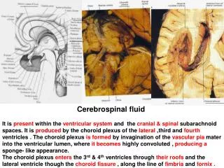
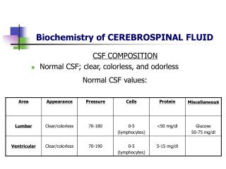
Cerebrospinal Fluid The cerebrospinal fluid is clear colorless fluid, contains a low amount of cells mainly lymphocytes. It is produced mainly by the choroid plexuses of the lateral ventricles, a small amount by the third and fourth ventricle. The total volume of the CSF in the ventricle system and the subarachnoid space varies from 80-150 mL, but the ventricular system alone contains 15-40 ml . Approx. 500 mL are produced during a 24 h period. • Produced in the lateral ventricle Interventricular foramen of Monroe third ventricle Aqueduct of Sylvius Fourth ventricle 1.Median aperture (foramen of Magendie) 2,3. lateral apererture (foramina Luschka) 2 1 3 Cerebellomedullary and pontine cisterns Subaracnhoid space Absorbed into venous system through the arachnoid villi lateral surfaces to the region of the superior saggital sinus Basal Cisterns
Thank you for attention GOOD LUCK FOR YOUR EXAMS Do not forget that anatomy is completely doable exam. The written is the difficult part in my opinion, it is stressful you have to be Concetrated as soon you pass the written and you have studied you will manage to pass the oral also. P.S.:Just relax because in the end everyone will…………
Good luck! Don’t forget to study the pituuehjdbndjry gland

![CEREBRAL CIRCULATION AND CEREBROSPINAL FLUID [CSF]](https://cdn2.slideserve.com/4005143/slide1-dt.jpg)