Rabies Virus
Use of rabies virus as a transneuronal tracer of neuronal connections: implications for the understanding of rabies pathogenesis Gabriella Ugolini NBCM, CNRS Gif-sur-Yvette. Kuypers & Ugolini, Trends Neurosci. 1990. Amplification of the signal: self-amplifying marker . 3°. Rabies


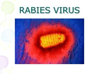
Rabies Virus
E N D
Presentation Transcript
Use of rabies virus as a transneuronal tracer of neuronal connections: implications for the understanding of rabies pathogenesis Gabriella Ugolini NBCM, CNRS Gif-sur-Yvette Kuypers & Ugolini, Trends Neurosci. 1990 Amplification of the signal: self-amplifying marker 3° Rabies Virus
Transneuronal tracing with rabies virus: 1 - Amplification of the signal: self-amplifying marker. 2 - Exclusive tropism for neurones in vivo. 3 - Absence of degeneration of infected neurones: possibility of combined visualisation of neurotransmitters & other tracers. 4 - Specificity: propagation exclusively by transneuronal transfer between connected neurones at chemical synapses. 5 - Intracellular transport is preferentially addressed to dendrites: transneuronal transfer occurs only in the retrograde direction. 6 - Ubiquitous distribution of rabies receptors in the CNS, but not in the peripheral nervous system. 7 -The only technique allowing the identification of neuronal connections across a practically unlimited number of synapses. CVS strain (1010 PFU/ml) Asymptomatic period
2 - No propagation via gap junctions (DM MNs) 3 - Sequential infection of 2°, 3 and 4° order neurons 4 - Centrifugal transfer to sensory and autonomic neurons at long time points, during the asymptomatic period 1 - Peripheral uptake is restricted to motoneurons: no uptake via sensory and autonomic neurons RAT Tang, Rampin, Giuliano & Ugolini (1999) J. Comp. Neurol. 414:167-192.
PRIMATES(macaque monkeys): injection into the lateral rectus (LR) muscle 1 - Peripheral uptake is restricted to motoneurons2 - Ubiquitous distribution of rabies receptors within the CNS3 - Centrifugal transfer to the vestibular (Scarpa’s) ganglionat 3 days p.i. (during the asymptomatic incubation period) Ugolini et al. J. Comp. Neurol. 2006
PRIMATES: differences in monosynaptic pathways to motoneurons of the lateral rectus muscle (LR) which innervate slow and fast muscle fibers • Muscle: defective replication in fibrocytes, no virus in myocites. • No spread within the muscle: uptake occurs only at the site of inoculation. • - Combined visualisation of rabies virus and choline acetyltransferase (CAT): infected motoneurons remain viable.
Primates: Injection of rabies virus into the CNS (Posterior parietal cortex, areas VIP, MIP) • Same pattern of propagation of rabies virus after central and peripheral inoculations: • 1 - Infection of first-order neurons at 2 days. • 2 - Transneuronal transfer occurs only retrogradely (no anterograde transfer to the pontine nuclei). • 3 - Transfer to connected neurons at sequential intervals of 12 hrs. • 4 - No local spread or cell death. 3° order at 3 days p.i.: centrifugal transfer to the vestibular (Scarpa’s) ganglion 2° order at 2.5 days p.i.: vestibular nuclei Co-injection of rabies virus & a conventional tracer (Cholera toxin B fragment, CTB) = no interference between uptake of rabies & CTB 3° Scarpa’s ganglion
Rabies pathogenesis: • In primates & rodents (rats, guinea pigs), peripheral uptake is restricted to motor endplates/axons: in keeping with the presynaptic location of NCAM and with a role of Ach nicotinic receptors (despite their mainly post-synaptic location). • Rabies virus does not spread within the muscle: uptake occurs only at the site of inoculation = importance of complete wound infiltration with rabies antibodies as soon as possible, to prevent virus entry! • Motoneurons are the only gateway for propagation of rabies virus to the CNS. • Rabies virus propagates exclusively by retrograde transneuronal transfer at chemical synapses - not via gap junctions or local spread. • Transneuronal transfer occurs only retrogradely – due to the fact that, after replication, centrifugal intracellular transport of rabies targets only dendrites, and not axons. • Retrograde transport and transneuronal transfer occur at high speed, by active axonal transport (P/LC8 interactions and microtubule-based transport) • Ubiquitous distribution of rabies receptor(s) within the CNS: transneuronal transfer involves all known populations connected to first-order neurons, regardless of their transmitters. • Extensive propagation of rabies virus within the CNS during the asymptomatic period! Each successive step of transfer (to 2°, 3°, 4° order neurons) requires only 12 hrs, regardless of the distance. • Infection of sensory and autonomic neurons requires longer incubation times because it reflects centrifugal propagation from the CNS to the periphery.
Acknowledgments Supported by: EU grants BIO4-CT98-0546 (TransVirus) & QLK6-CT-2002-0015 (EUROKINESIS)