Download
1 / 60
600 likes | 734 Views
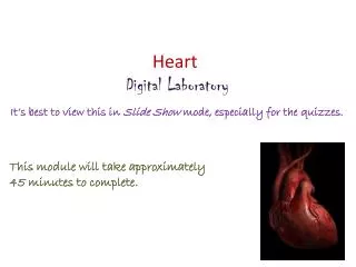
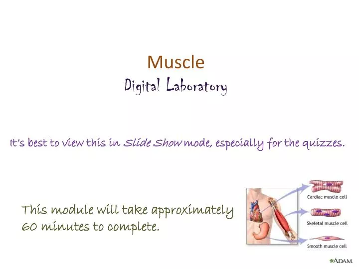
Muscle Digital Laboratory. It’s best to view this in Slide Show mode, especially for the quizzes. This module will take approximately 60 minutes to complete. After completing this exercise, you should be able to: Distinguish, at the light microscope level, each of the following: Muscle

E N D
Muscle Digital Laboratory It’s best to view this in Slide Show mode, especially for the quizzes. This module will take approximately 60 minutes to complete.
After completing this exercise, you should be able to: • Distinguish, at the light microscope level, each of the following: • Muscle • Skeletal muscle • Cardiac muscle • Intercalated disks • Smooth muscle • Distinguish, at the electron microscope level, each of the following: • Smooth muscle • Myofilaments (mostly actin filaments) • Dense bodies • Caveolae (pinocytotic vesicles) • Gap junctions
MUSCLE TISSUE Muscle tissue has one common function: it shortens (contracts). This is accomplished by muscle cells, which are held together by connective tissues that also contain the blood vessels and nerves that support the muscle cells. There are three types of muscle tissue: Skeletal muscle is involved in voluntary contraction, and is associated with the body wall. This is the tissue of your named muscles (biceps brachii, petoralis major) Cardiac muscle is found in the heart (FYI and pulmonary veins near the heart). Obviously it is involuntary. Smooth muscle is found in visceral organs, such as the gastrointestinal, respiratory, urinary, and reproductive systems. It is also found in the body wall, specifically smooth muscle of blood vessels as well as arrector pili muscles of the hair follicles. This module will focus on recognizing muscle tissue, and differentiating between the three types of muscle. It will then explore smooth muscle in a little more detail, because smooth muscle is ubiquitous. Skeletal muscle will be examined in greater detail in the Musculoskeletal and Integument Block, and cardiac muscle will be revisited in the Cardiovascular, Renal, and Pulmonary Block.
SKELETAL MUSCLE Muscles like the biceps brachii are composed of skeletal muscle cells bundled in connective tissue sheaths; this organization is similar to the bundling of axons in nerves. The details of the organization of the sheaths is not relevant here. What is important to appreciate now is that skeletal muscle cells are very large, both in length and diameter, and are called muscle fibers. In a muscle, the muscle fibers are all arranged in the same orientation. Also note that each muscle fiber (cell) is packed with longitudinal structures called myofibrils, which are composed of contractile proteins.
Formation of a skeletal muscle fiber (muscle cell) A longitudinal section of skeletal muscle like the one shown above will have a characteristic dark-light banding pattern. However, also note that a good, high magnification view of skeletal muscle in cross section will show stippling within the cell due to sectioning of the myofibrils. Skeletal muscle cells (fibers) develop from the fusion of myoblasts, resulting in large, multinuclear cells (each cell is a syncytium – how cool is that). The cells then assemble their contractile machinery in the cytoplasm. These come in the form of myofibrils, which have an alternate dark-light banding pattern when viewed from the side. The fact that the cell is chock-full of these myofibrils pushes the nuclei to the periphery of the cell.
SKELETAL MUSCLE Here are two images from our slide set, taken at medium and very high magnification (oil). Both are longitudinal views of skeletal muscle. The muscle fibers (cells) are indicated by the brackets. Typically, within a single muscle, all fibers are the same diameter, so the apparent difference you see is due to sectioning (e.g. the section goes through the middle of some fibers, cuts the edge of others). Note the fact that these are long, wide-diameter cells (compare to the size of the nuclei), with an alternating dark-light pattern, with most nuclei situated in the periphery of the cell. Actually, some nuclei belong to the muscle cells, while others are of fibroblasts. Difficult to tell for sure, but the muscle nucleus is typically more euchromatic than the fibroblast nucleus, so I’m going with the nucleus indicated by the black arrow belonging to the muscle cell, and the one indicated by the blue arrow belonging to a fibroblast that is in the loose connective tissue between cells. Just an educated guess. Nothing to worry about now.
SKELETAL MUSCLE Video of skeletal muscle – SL86 • Link to SL 086 • Be able to identify: • Skeletal muscle
SKELETAL MUSCLE The previous slide was a plastic section, and oriented so the muscle fibers were all cut longitudinally. Like real life, the rest of our slides aren’t so perfect. Here you have a typical longitudinal view of skeletal muscle. The diameter of a single fiber (cell) is indicated by the brackets. Note the intense cytoplasmic eosinophilia, caused by the tremendous amount of contractile proteins in these cells. Here you can still see striations, and that most nuclei are toward the periphery of the cell. However, cell borders are not as obvious; in fact, it’s actually the nuclei that help you “see” the cell borders. The nuclei are elongated, but plump, like a bratwurst (image supplied to the right for those not native to Cincinnati).
SKELETAL MUSCLE Life is even less fair. The previous image was selected because it was a region in which the cells were at a perfect longitudinal angle. Turning the angle ever so slightly eliminates the striations you would like to rely on. Here you can still see that the diameter of a cell is wide (yellow brackets), based on the positioning of the nuclei and the loose connective tissue that separates the cells. Fortunately, you should have the option of scanning around the slide to find nice striations – but no guarantees.
SKELETAL MUSCLE In the same slide (and others in our set), some of the fibers are oriented in cross-section; you need to be able to recognize skeletal muscle in cross-section as well. In cross sections, usually it is easier to see the cell borders and peripheral nuclei. Also, if you look closely, you can see stippling within the cytoplasm (outlined cell is best for this), which represents the myofibrils cut in cross section.
SKELETAL MUSCLE Alas, like longitudinal sections, perfect cross-sections are not always the norm. Here, even a slight angle takes away the obvious stippling (some short “bands” may be visible), and cell borders (brackets indicate cells) are not as distinct.
SKELETAL MUSCLE Video of skeletal muscle – SL27 • Link to SL 027 and SL 061 and SL 060 • Be able to identify: • Skeletal muscle
CARDIAC MUSCLE Cardiac muscle is composed of smaller, branched muscle cells, which are connected to each other by intercalated discs. These intercalated disks, which are unique to cardiac muscle tissue, include adherent junctions for cell-cell strength, as well as gap junctions to allow electrical synchrony (so the cells contract at the same time). Similar to skeletal muscle, cardiac muscle fibers are packed with myofibrils, which are in-register, and give the tissue a striated appearance. Each cardiac muscle cell has a single nucleus that is centrally located. When we say “smaller cells” for cardiac muscle, this is a comparison to skeletal muscle cells. Turns out, cardiac muscle cells are quite large when compared to most other cells, including smooth muscle cells we’ll see later. They just happen to be smaller than the very large skeletal muscle cells.
CARDIAC MUSCLE Just like skeletal muscle, striations are readily apparent in cardiac muscle when viewed perpendicular to the orientation of the cells. Also like skeletal muscle, the fibers are long, with a consistent diameter throughout the length of the cell (green brackets). The diameter of each cell is similar, the slight variation due to sectioning (either through the thickest part of the cell, or catching the edge). However, the diameter of each cell (brackets) is much narrower than skeletal muscle. This image gives you the impression that the diameter of each cell is comparable to the size of the nucleus. This is not the case; in fact, cardiac muscle cells are considered to have a fairly wide diameter relative to most cells (though much smaller than skeletal muscle). This will be better seen in cross-section.
CARDIAC MUSCLE Note also the centrally-located nuclei, and one per cell (though the later statement is hard to see). The nuclei here appear perfectly round, though typically they are oval, just not as elongated as seen in skeletal muscle. The ends of the cells are joined by intercalated disks (black arrows), which appear as dense bands in the same orientation as the striations. The cells are also branched, a nice example of a branched cell is in the insert in the upper left.
CARDIAC MUSCLE This image is a cross-section through cardiac muscle tissue. Three cells are outlined, color coded to the section they represent in the cartoon to the right. You can see the centrally-located nuclei, although the nucleus is only visible in cells sectioned through the nucleus (black), while in other cells the nucleus is not in the same plane as the section (yellow). Like skeletal muscle, cardiac muscle cells have approximately the same diameter throughout their length. Therefore, all cells have approximately the same diameter. Cells with oval shapes (purple) are due to sectioning through branch points. The relatively large rim of cytoplasm around the nucleus will become useful when comparing to smooth muscle.
CARDIAC MUSCLE Video of cardiac muscle – SL88 • Link to SL 088 • Be able to identify: • Cardiac muscle • Intercalated disks
CARDIAC MUSCLE In this specially-stained slide (Bencosme), intercalated disks are easier to see (arrows).
CARDIAC MUSCLE Video of cardiac muscle showing intercalated disks – SL64 • Link to SL 064 • Be able to identify: • Cardiac muscle • Intercalated disks
CARDIAC MUSCLE A scanning electron micrograph (a) was taken from cardiac muscle specially prepared to remove connective tissue elements and separate cardiac muscle cells at their intercalated disks. The end of a cell is outlined. A cartoon (b) is labeled. In the transmission electron micrograph (c), cells run longitudinally from upper left to lower right. A dotted red line (visible when you advance the slide) indicates an intercalated disk, which joins the cells “end-to-end”. Fascia adherens (FA) and macula adherens (MA) anchor the ends of cells together, while gap junctions (GJ), which are only on the “sides” of the disk, provide electrical continuity between the cells. Fascia adherens is similar to the zonulaadherens of epithelial cells.
SMOOTH MUSCLE Smooth muscle tissue is composed of many smooth muscle cells. Although there are connective tissue elements (e.g. collagen) between the cells, smooth muscle is much more cellular than connective tissue. In addition, smooth muscle cells are smaller than cardiac and skeletal muscle cells. These features result is a tissue that has lots of nuclei. Depending on the orientation of the cells, the nuclei are slightly elongated in longitudinally-oriented cells (L), or round in transverse (cross) -sections (T). The contractile proteins within smooth muscle cells give the tissue a highly eosinophilic color, though usually (not always) less eosinophilic that cardiac or skeletal muscle.
SMOOTH MUSCLE Here’s a nice slide that is a unique view of smooth muscle because individual smooth muscle cells (outlined) are well-defined. As we will see in subsequent slides, this in almost never the case. Note that the cells are small (actually, their size is typical as far as cells go, but small when compared to skeletal and cardiac muscle). They are tapered or cigar shape, so they are thinner at the tips and fatter in the middle, where a single, central nucleus resides (arrows). In this middle portion of the cell, there is relatively little cytoplasm surrounding the nucleus. Loose connective tissue is between the cells. Yep, looks a little striated in places. This is due to pixilation, trust me. from Wheater’s Functional Histology
SMOOTH MUSCLE This image is more typical of smooth muscle because it is difficult to see individual cells. Note that: --The tissue has an overall appearance that is more red in color than other tissues, but typically is slightly pinker (less red) than skeletal or cardiac muscle. --Note the oval shaped nuclei. Also note that the nuclei are relatively evenly dispersed throughout the tissue. Connective tissues tend to have fewer nuclei that are more clustered or unevenly distributed. --The contractile proteins are NOT arranged in an ordered fashion, thus there are no visible striations in smooth muscle. I know, there are “striations” here, but I swear, this is an artifact, and they usually aren’t there (and will NOT be present on an exam slide of smooth muscle).
SMOOTH MUSCLE This image is similar to the one three slides previous. The left portion of the image is a longitudinal view of smooth muscle, while the right side is a cross-section. Once again, cell borders are difficult to discern. It can be done with high magnification, as we will see shortly. However, some key features to note: 1. Numerous nuclei due to the fact that there are many, small cells 2. Eosinophilia due to abundant contractile proteins in the cytoplasm of these cells 3. In longitudinal sections, the nuclei are oval; in cross section, the nuclei are round 4. There is no distinct “border” around the smooth muscle, it blends in with the surrounding connective tissue. This is undoubtedly subtle, and is less clear because you have yet to study the pale structure in the center of this image, which is a ganglion (part of the nervous system) and does have a distinct border. Information provided on organs in this group of smooth muscle images is FYI for now colon
SMOOTH MUSCLE A survey of smooth muscle for your viewing pleasure…. Smooth muscle often comes in bundles. Here, bundles of smooth muscle (outlined) are surrounded by dense irregular connective tissue. Note more nuclei in smooth muscle when compared to the surrounding connective tissue. Section of nipple
SMOOTH MUSCLE A survey of smooth muscle for your viewing pleasure…. More bundles of smooth muscle surrounded by connective tissue. Cross section is the top bundle, longitudinal section is below. urinary bladder
SMOOTH MUSCLE A survey of smooth muscle for your viewing pleasure…. Here, the bundles are intertwined Uterus (myometrium)
SMOOTH MUSCLE A survey of smooth muscle for your viewing pleasure…. In images of cross sections of smooth muscle taken at higher magnification, various cross-sectional profiles of smooth muscle cells are visible. Sections through the center of the cell (green) show nuclei, are largest in diameter, with a central nucleus. Sections through the periphery of the cell (blue) lack a nucleus, and vary in diameter.
SMOOTH MUSCLE Video of urinary bladder showing the smooth muscle – SL57 • Link to SL 057 and SL 053 and SL 138and SL 155 • Be able to identify: • Smooth muscle
SMOOTH MUSCLE All muscle cell shortening occurs due to the action of the contractile proteins actin and myosin. You will learn the organization of skeletal muscle proteins, as well as their mechanism of action, in greater detail in the Musculoskeletal block. Here, we will briefly introduce smooth muscle ultrastructure. In smooth muscle, actin (microfilaments) are more predominant than myosin. In the image to the left, portions of three smooth muscle cells are shown (one is outlined). Note that they are packed with fine microfilaments. Other characteristics of smooth muscle cells include caveoli or pinocytotic vesicles (PV), and dense bodies (arrows), into which the actin filaments are anchored, and gap junctions (GJ) for intercellular communication.
SMOOTH MUSCLE The inset in the upper left shows about 6 smooth muscle cells. The larger image is similar to the boxed region in the inset. Again note that the cytoplasm of the cells is filled with microfilaments. Arrows indicate dense bodies; the double arrows show larger dense bodies where they anchor to the plasma membrane. PV = pinocytotic vesicles; BL = basal lamina
SKELETAL MUSCLE & SMOOTH MUSCLE In real, real life, or at least practical exam real life, you will have to distinguish skeletal muscle from smooth muscle, and connective tissue. Fortunately for you, we have just the slide. This is a section of the esophagus, in a region of transition from skeletal (voluntary) muscle to smooth (autonomic) muscle. Skeletal muscle is typically more eosinophilic (on the red side, as opposed to pink), with large-diameter cells, peripheral nuclei. Smooth muscle cells are smaller, so more nuclei, evenly distributed. On a much more subtle note, the nuclei are more heterochromatic than those of skeletal muscle, and some even look “twisted” (arrow). Dense irregular connective tissue has fewer cells, so fewer nuclei, with extracellular elements such as collagen fibers. Smooth muscle Dense irregular c.t. Skeletal muscle
SKELETAL MUSCLE & SMOOTH MUSCLE Same slide of the esophagus, most of the fibers here are in cross-section. Skeletal muscle is typically more eosinophilic, with large-diameter cells, peripheral nuclei. Smooth muscle cells are smaller, so more nuclei, relatively evenly distributed. The increase in the number of nuclei is not so obvious in cross-section, but if you look closely, you can see individual cells, some with central nuclei and a small rim of cytoplasm (blue arrows), others are smaller in diameter without nuclei representing tapered ends of cells (black arrows). I guesstimate that there are hundreds of cells in the upper half of the outlined region. (Arrows on peripheral cells so as not to obscure your view.) Dense irregular connective tissue has fewer cells, so fewer nuclei, with extracellular elements such as collagen fibers. Skeletal muscle Dense irregular c.t. Smooth muscle
SKELETAL MUSCLE & SMOOTH MUSCLE Video of skeletal muscle and smooth muscle – SL15A • Link to SL 015A • Be able to identify: • Skeletal muscle • Smooth muscle • Dense irregular connective tissue (review)
COMPARISON OF SKELETAL, CARDIAC, & SMOOTH MUSCLE First off, for all you “memorizers” out there, here’s a list of features of each tissue type: Characteristics of skeletal muscle cells: -Long cells -fibers of constant diameter, wide diameter -Multiple nuclei, located in periphery of cell -Striated -Voluntary -Found in named muscles (e.g. biceps) Characteristics of cardiac muscle cells: -longer than typical cells, though much shorter than skeletal muscle cells -fibers of constant diameter, intermediate diameter -Central nucleus -Striated -Inoluntary -Found in heart -Branched cells -Intercalated disks Characteristics of smooth muscle cells: -Spindle-shaped cells (cigar-shaped) -Central nucleus -Not striated -Involuntary -Found in visceral organs, blood vessels
COMPARISON OF SKELETAL, CARDIAC, & SMOOTH MUSCLE --Skeletal and cardiac muscle tend more toward the red shade, while smooth muscle is pink (not absolute). --Striations are evident in skeletal and cardiac, but not smooth. --Nuclei are located in the center of each cardiac and smooth muscle cell, but are located near the plasma membrane in skeletal muscle. --Skeletal muscle cells are large and multinuclear, while cardiac and smooth muscle cells are smaller, with only a single nucleus. --Because smooth muscle cells are smaller than either cardiac or skeletal muscle cells, smooth muscle typically has more visible nuclei than the other two tissues. This is not obvious on this example. --Cardiac muscle is the only tissue that has intercalated disks. cardiac muscle 480X skeletal muscle 320X smooth muscle 320X
COMPARISON OF SKELETAL, CARDIAC, & SMOOTH MUSCLE --Skeletal muscle cells have the largest diameter, cardiac muscle cells are smaller, and smooth muscle cells have the smallest diameter. --The diameter of skeletal muscle is very consistent between cells. Cardiac muscle cells are somewhat consistent in diameter (but vary in shape), while the smooth muscle cell profiles vary depending on whether the cell is sectioned through the middle (with nucleus) or near the periphery of the cell (without nucleus). --Skeletal muscle nuclei are near the plasma membrane, while cardiac and smooth muscle nuclei are centrally located. --In cuts through the nucleus of a cell, there is significant cytoplasm in skeletal and, to a lesser extent, cardiac muscle, while smooth muscle has a small rim of cytoplasm around the nucleus. --In ideal sections, stippling can be seen in skeletal and cardiac muscle, but not smooth muscle. skeletal muscle 320X cardiac muscle 480X smooth muscle 320X
The next set of slides is a quiz for this module. You should review the structures covered in this module, and try to visualize each of these in light and electron micrographs. • Distinguish, at the light microscope level, each of the following: • Muscle • Skeletal muscle • Cardiac muscle • Intercalated disks • Smooth muscle • Distinguish, at the electron microscope level, each of the following components of connective tissue: • Smooth muscle • Myofilaments (mostly actin filaments) • Dense bodies • Caveolae (pinocytotic vesicles) • Gap junctions
Final quiz Self-check: Identify the tissue. (advance slide for answers) Smooth muscle Smooth muscle
Final quiz Self-check: Identify the tissue in the outlined region. (advance slide for answers) Smooth muscle
Final quiz Self-check: Identify the outlined tissues. (advance slide for answers) Smooth muscle Dense irregular connective tissue
Final quiz Self-check: Identify the outlined TISSUE. (advance slide for answers) Skeletal muscle
Final quiz Self-check: Identify the outlined TISSUES. (advance slide for answers) Dense irregular connective tissue Smooth muscle
Final quiz Self-check: Identify structure indicated by the arrows. (advance slide for answers) Intercalated disk
Final quiz Self-check: Identify the outlined TISSUES. (advance slide for answers) Dense irregular connective tissue (elastic tissue) Skeletal muscle
Final quiz Self-check: Identify the tissue. (advance slide for answers) cardiac muscle
Final quiz Self-check: Identify the tissue. (advance slide for answers) skeletal muscle
Final quiz Self-check: Identify the tissue. (advance slide for answers) skeletal muscle
Final quiz Self-check: Identify the epithelium on this slide. (advance slide for answer) transitional
Final quiz Self-check: Identify the tissue from which this micrograph was taken. (advance slide for answers) smooth muscle microfilaments Pinocytotic vesicles