Download
1 / 95
970 likes | 1.22k Views
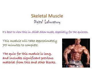
Skeletal Muscle Digital Laboratory. It’s best to view this in Slide Show mode, especially for the quizzes. This module will take approximately 75 minutes to complete. The quiz for this module is long, and includes significant previous material from this and other blocks. Lowrie.

E N D
Skeletal Muscle Digital Laboratory It’s best to view this in Slide Show mode, especially for the quizzes. This module will take approximately 75 minutes to complete. The quiz for this module is long, and includes significant previous material from this and other blocks. Lowrie
After completing this exercise, you should be able to: • identify, at the light microscope level, each of the following: • Skeletal muscle • Myofibers (muscle fibers, muscle cells) • Myofibrils • Fascicle • Endomysium • Perimysium • Epimysium • Bands and lines seen in skeletal muscle • A band • I band • H band • Z line • M line • Sarcomere • identify, at the electron microscope level, each of the following: • Skeletal muscle • Myofibers (muscle fibers, muscle cells) • Myofibrils • Myofilaments • Thick filaments • Thin filaments • Sarcolemma • Bands and lines noted above for light microscopy • Glycogen and Mitochondria • FYI… • (Sarcoplasmic reticulum) • (Triad) • (Terminal cisternae of sarcoplasmic reticulum (x2)) • (Transverse tubule (T-tubule) (x1))
SKELETAL MUSCLE & SMOOTH MUSCLE Recall that in the Fundamentals block, we had mini-modules on skeletal and smooth muscle so that you could recognize these tissues on slides from that block. The next 12 slides are from the portion describing skeletal muscle and comparing it to smooth muscle and dense irregular connective tissue. I recommend that you review these now. However, if you’re absolutely sure you can recognize these no problem in light micrographs and slides (I have provided examples for your viewing pleasure), you can click here to skip them. Otherwise, proceed as normally. Hint: If you are hoping for answers to what you’re looking at, then you aren’t 100% certain, and you probably should go through the slides. FYI, a technical note: When looking at these slides now, I had the notion to make minor changes in them to fit the module that I am creating for this block. However, if I did so, then everyone would feel they had to go through them to see what I changed. Therefore, I am leaving these 12 slides completely intact as they were. Because of this, some of the statements may seem out of context for today’s assignment.
SKELETAL MUSCLE Muscles like the biceps brachii are composed of skeletal muscle cells bundled in connective tissue sheaths; this organization is similar to the bundling of axons in nerves. The details of the organization of the sheaths is not relevant here. What is important to appreciate now is that skeletal muscle cells are very large, both in length and diameter, and are called muscle fibers. In a muscle, the muscle fibers are all arranged in the same orientation. Also note that each muscle fiber (cell) is packed with longitudinal structures called myofibrils, which are composed of contractile proteins.
Formation of a skeletal muscle fiber (muscle cell) A longitudinal section of skeletal muscle like the one shown above will have a characteristic dark-light banding pattern. However, also note that a good, high magnification view of skeletal muscle in cross section will show stippling within the cell due to sectioning of the myofibrils. Skeletal muscle cells (fibers) develop from the fusion of myoblasts, resulting in large, multinuclear cells (each cell is a syncytium – how cool is that). The cells then assemble their contractile machinery in the cytoplasm. These come in the form of myofibrils, which have an alternate dark-light banding pattern when viewed from the side. The fact that the cell is chock-full of these myofibrils pushes the nuclei to the periphery of the cell.
SKELETAL MUSCLE Here are two images from our slide set, taken at medium and very high magnification (oil). Both are longitudinal views of skeletal muscle. The muscle fibers (cells) are indicated by the brackets. Typically, within a single muscle, all fibers are the same diameter, so the apparent difference you see is due to sectioning (e.g. the section goes through the middle of some fibers, cuts the edge of others). Note the fact that these are long, wide-diameter cells (compare to the size of the nuclei), with an alternating dark-light pattern, with most nuclei situated in the periphery of the cell. Actually, some nuclei belong to the muscle cells, while others are of fibroblasts. Difficult to tell for sure, but the muscle nucleus is typically more euchromatic than the fibroblast nucleus, so I’m going with the nucleus indicated by the black arrow belonging to the muscle cell, and the one indicated by the blue arrow belonging to a fibroblast that is in the loose connective tissue between cells. Just an educated guess. Nothing to worry about now.
SKELETAL MUSCLE Video of skeletal muscle – SL86 • Link to SL 086 • Be able to identify: • Skeletal muscle
SKELETAL MUSCLE The previous slide was a plastic section, and oriented so the muscle fibers were all cut longitudinally. Like real life, the rest of our slides aren’t so perfect. Here you have a typical longitudinal view of skeletal muscle. The diameter of a single fiber (cell) is indicated by the brackets. Note the intense cytoplasmic eosinophilia, caused by the tremendous amount of contractile proteins in these cells. Here you can still see striations, and that most nuclei are toward the periphery of the cell. However, cell borders are not as obvious; in fact, it’s actually the nuclei that help you “see” the cell borders. The nuclei are elongated, but plump, like a bratwurst.
SKELETAL MUSCLE Life is even less fair. The previous image was selected because it was a region in which the cells were at a perfect longitudinal angle. Turning the angle ever so slightly eliminates the striations you would like to rely on. Here you can still see that the diameter of a cell is wide (yellow brackets), based on the positioning of the nuclei and the loose connective tissue that separates the cells. Fortunately, you should have the option of scanning around the slide to find nice striations – but no guarantees.
SKELETAL MUSCLE In the same slide (and others in our set), some of the fibers are oriented in cross-section; so, like smooth muscle, you need to be able to recognize skeletal muscle in cross-section as well. In cross sections, usually it is easier to see the cell borders and peripheral nuclei. Also, if you look closely, you can see stippling within the cytoplasm (outlined cell is best for this), which represents the myofibrils cut in cross section.
SKELETAL MUSCLE Alas, like longitudinal sections, perfect cross-sections are not always the norm. Here, even a slight angle takes away the obvious stippling (some short “bands” may be visible), and cell borders (brackets indicate cells) are not as distinct.
SKELETAL MUSCLE Video of skeletal muscle – SL27 • Link to SL 027and SL 061and SL 060 • Be able to identify: • Skeletal muscle
SKELETAL MUSCLE & SMOOTH MUSCLE And in real, real life, or at least practical exam real life, you will have to distinguish skeletal muscle from smooth muscle, and connective tissue. Fortunately for you, we have just the slide. This is a section of the esophagus, in a region of transition from skeletal (voluntary) muscle to smooth (autonomic) muscle. Skeletal muscle is typically more eosinophilic (on the red side, as opposed to pink), with large-diameter cells, peripheral nuclei. Smooth muscle cells are smaller, so more nuclei, evenly distributed. On a more subtle note, the nuclei are more heterochromatic than those of skeletal muscle, and some even look “twisted” (arrow). Dense irregular connective tissue has fewer cells, so fewer nuclei, with extracellular elements such as collagen fibers. Smooth muscle Dense irregular c.t. Skeletal muscle
SKELETAL MUSCLE & SMOOTH MUSCLE Same slide of the esophagus, most of the fibers here are in cross-section. Skeletal muscle is typically more eosinophilic, with large-diameter cells, peripheral nuclei. Smooth muscle cells are smaller, so more nuclei, relatively evenly distributed. The increase in the number of nuclei is not so obvious in cross-section, but if you look closely, you can see individual cells, some with central nuclei and a small rim of cytoplasm (blue arrows), others are smaller in diameter without nuclei representing tapered ends of cells (black arrows). I guesstimate that there are hundreds of cells in the upper half of the outlined region. (Arrows on peripheral cells so as not to obscure your view.) Dense irregular connective tissue has fewer cells, so fewer nuclei, with extracellular elements such as collagen fibers. Skeletal muscle Dense irregular c.t. Smooth muscle
SKELETAL MUSCLE & SMOOTH MUSCLE Video of skeletal muscle and smooth muscle – SL15A • Link to SL 015A • Be able to identify: • Skeletal muscle • Smooth muscle (review) • Dense irregular connective tissue (review)
SKELETAL MUSCLE - ORGANIZATION Recall this drawing of a muscle such as the rectus femoris. A muscle is composed of muscle cells, called muscle fibers, or myofibers, all running parallel within the muscle. Within a muscle, a number of muscle fibers are bundled together into fascicles (dotted outline). The connective tissue component of a muscle can be divided into three types: Endomysium – between individual muscle fibers Perimysium – around fascicles Epimysium – surrounds the entire muscle The epimysium is the deep fascia that surrounds muscles in the gross anatomy lab. Consistent with connective tissue organization in other organs you have seen, the density of the connective tissue decreases as you progress toward the inner portions of the muscle. In other words, epimysium is dense irregular connective tissue, endomysium is loose connective tissue, and perimysium is somewhere in between (the famous “loosy-densy” connective tissue).
SKELETAL MUSCLE - ORGANIZATION In this low power image of an entire muscle, a fascicle is outlined, and the epimysium (black arrows) and perimysium (blue arrow) are indicated. The epimysium is the deep fascia that surrounds muscles in the gross anatomy lab.
SKELETAL MUSCLE - ORGANIZATION This higher powered micrograph from the edge of a muscle shows a portion of a fascicle; a single muscle fiber is outlined. The epimysium (black arrow), perimysium (blue arrow) and endomysium (green arrow) are indicated. Apart from the difference in density, these layers really don’t look much different. They are all irregular connective tissues, either loose, dense, or something in between. They are named based on their position in the muscle and among the muscle fibers.
SKELETAL MUSCLE - ORGANIZATION Video of skeletal muscle showing epimysium, perimysium, endomysium – SL60 • Link to SL 060 • Be able to identify: • Skeletal muscle • Fascicle • Endomysium • Perimysium • Endomysium
SKELETAL MUSCLE CELLS As we mentioned, skeletal muscle cells are very large cells, both in diameter and length, and are, therefore, called muscle fibers, or myofibers. Their cytoplasm is packed with contractile proteins, which are organized into rod-like structure called myofibrils. Like neuroscientists, people who study muscle have a big ego. Therefore, they rename all the cellular components with their “sarco” prefix, presumably because these components in muscles are better than those same components in the liver or bone. Therefore: sarcolemma = plasmalemma sarcoplasm = cytoplasm sarcoplasmic reticulum = (smooth) endoplasmic reticulum Because in skeletal muscle we have rods within rods within rods, it’s easy to lose perspective and get lost in the terminology. Don’t forget that each part of the drawing here (top and bottom) shows a single muscle cell.
SKELETAL MUSCLE CELLS In the same slide (and others in our set), some of the fibers are oriented in cross-section. As is the case for smooth muscle, you need to be able to recognize skeletal muscle in cross-section. A single muscle cell is outlined, two more are indicated by the green brackets. In cross sections, usually it is easier to see the cell borders and peripheral nuclei. Also, if you look closely, you can see stippling within the cytoplasm (outlined cell is best for this), which represents the myofibrils cut in cross section. No sense recreating the wheel. This is a slide from the previous module with the inset added. The region within the box has been artificially magnified, so I could put a few arrows on three of the hundreds of cross sections of myofibrils. Saaaweeeet.
SKELETAL MUSCLE CELLS Video of skeletal muscle showing myofibrils in cross section – SL60 • Link to SL 027 • Be able to identify: • Skeletal muscle • Myofibers • Myofibrils
SKELETAL MUSCLE CELLS The image on the left is a longitudinal section of about 6-8 skeletal muscle fibers (cells); the green bracket indicates one cell. The dark-light-dark-light-etc. banding pattern you see, which will be explained in detail on the next set of slides, is created by the fact that each myofibril in a cell has a dark-light-dark-light-etc. banding pattern, and the myofibrils are arranged so that their dark bands line up side-by-side, as do the light bands. TOTALLY AWESOME!!!! Some 30-50 or so myofibrils span the diameter of a single cell. Because they are “in register”, you can’t see most myofibrils. However, during fixation, some of them “slide”, putting them out of register with their neighbors. Between the arrows is probably a single myofibril that has slid, giving you an idea of the diameter of a single myofibril.
SKELETAL MUSCLE CELLS Video of skeletal muscle showing myofibrils in longitudinal section – SL86 • Link to SL 086 • Be able to identify: • Skeletal muscle • Myofibers • Myofibrils
SKELETAL MUSCLE CELLS – BANDING PATTERN The banding pattern you see in light micrographs of skeletal muscle is created by the collective banding of each myofibril (e.g. between yellow arrows). A bands (green bracket) and I bands (black bracket) are dark and light, respectively. Also readily seen is the dark line within the I band, the Z line (black arrows). Less obvious is a pale region within the A band, referred to as the H band or H zone (maroon arrows). The functional contractile unit of skeletal muscle, the scaromere, extends from Z line to Z line (blue bracket)
SKELETAL MUSCLE CELLS – BANDING PATTERN Video of skeletal muscle showing banding pattern – SL86 • Link to SL 086 • Be able to identify: • Skeletal muscle • A band • I band • Z line • H band (H zone) • sarcomere
SKELETAL MUSCLE CELLS – BANDING PATTERN Moving into the World of EMs. Not much to say here, just showing you an EM (right) of comparable magnification to the light micrographs you have been looking at recently. Longitudinal axis of muscle cell Width of muscle cell Nuclei of muscle cell Endomysium Capillaries Striations
SKELETAL MUSCLE CELLS – BANDING PATTERN This EM was taken at slightly higher magnification than the previous slide. The width of a single muscle cell is indicated by the red double-arrow. Note the A and I bands, as well as the Z line and H zone (later between yellow arrows) Capillary lumen Muscle cell nuclei Endothelial cell nucleus (capillary-lining cell) Endomysium A bands I bands (lines actually touching Z lines) Longitudinal axis of cell
SKELETAL MUSCLE CELLS – BANDING PATTERN Enlargement of a single sarcomere is shown to the right. The extent of a single sarcomere is indicated by the purple double-arrow. Full labeling on next slide…
SKELETAL MUSCLE CELLS – BANDING PATTERN Z line I band (note single I band extends on both sides of Z line) Border between A and I band A band (actually, 4,5,6, and 7 are all in A band) Ignore this for now H zone Ignore the rest of these for now Note that adjacent sarcomeres share a Z line. Also, as indicated in #2 above, a single I band consists of parts of two adjacent sarcomeres. Take note of the filamentous proteins that run along the longitudinal axis of the sarcomere in this image. These are the myofilaments, and will be discussed in the next set of slides.
SKELETAL MUSCLE CELLS – MYOFILAMENTS There are two types of myofilaments: Thin filaments – made up of actin (yes, the cytoskeletal element called microfilaments) Thick filaments – made up of myosin (neither microtubules nor intermediate filaments) As you can see in the schematic drawing and electron micrograph, myofilaments are oriented along the long axis of the myofibrils (and myofibers). Each myofilament is anchored by a collection of proteins that form specific lines: The Z line anchors the thin filaments The M line anchors the thick filaments Or, from a different perspective, the Z line has numerous thin filaments extending from it in both directions, and the M line has numerous thick filaments extending from it in both directions. Also note that thin and thick filaments extend past each other, creating a zone of overlap.
SKELETAL MUSCLE CELLS – MYOFILAMENTS It should also be pointed out that the myofibrils are cylindrical, not flat structures. Therefore, although we use the terms Z lines and M lines when looking at longitudinal sections of muscle, these structures are actually disk shaped, and are often called Z disks or M disks. Also, they do have some thickness, though they are very thin, so they are really puck-shaped.
SKELETAL MUSCLE CELLS – MYOFILAMENTS Just to be sure, in this enlarged EM: Thin filaments (orange arrows) – made up of actin (hey, these are thin, but several look a little chubby) Thick filaments (purple arrows) – made up of myosin Only a few examples are pointed out here. If you start at the blue block arrow and work upward, you probably will encounter 20-30 thick filaments.
SKELETAL MUSCLE CELLS – BANDING PATTERN Now we can identify more stuff…. Z line I band (note it extends on both sides of Z line) Border between A and I band A band (actually, 4,5,6, and 7 are all in A band) Ends of thin filaments H zone M line Extent of thin filaments Extent of thick filaments 10-14. Ignore these for now Note the zone of overlap as well (4), the darkest region of the A band, except maybe the M line. You will learn more about the detailed structure and function of these myofilaments in your reading assignment and large-group sessions.
SKELETAL MUSCLE CELLS – BANDING PATTERN To solidify your understanding, you probably want to think about what cross-sections through each region would look like. This is purely FYI, since I won’t put cross-sectional EMs on an exam (though I have been known to put glass slides of skeletal muscle in cross section on exams). However, this exercise should help you remember the structure on longitudinal sections. Zone of overlap – note both thick (17) and thin (16) filaments M line – note only thick filaments (17); proteins linking thick filaments are slightly visible (arrows) H zone – only thick filaments (without proteins that bind them, though I can’t deny that it sure looks like some are there)
SKELETAL MUSCLE CELLS – BANDING PATTERN I band – thin filaments (16) only (12 is glycogen) Z line – thin filaments (16) cross linked by Z line proteins
SKELETAL MUSCLE CELLS – TUBULE SYSTEM As you will learn in the large-group sessions, muscle contraction requires calcium. The large diameter of skeletal muscle requires an elaborate cellular architecture to ensure that all myofibrils receive this calcium signal at the same time. This architecture has two major components: Invaginations of the sarcolemma (plasma membrane) called T tubules. Action potentials that travel along the surface sarcolemma use the T tubule system to bring the excitation wave to the center of the cell. The sarcoplasmic reticulum (endoplasmic reticulum) is elaborate, highlighted by dilations called terminal cisterna, which flank the T tubules. The sarcoplasmic reticulum stores calcium, which is released when calcium channels in the bilayer of the reticulum are exposed to action potentials travelling down the T tubules. The three tubules together, a central T tubule flanked by two terminal cisternae, form a “triad”. Human skeletal muscle has a “triad”, while human cardiac muscle (and muscle in other animals) has only one terminal cisternae, and, therefore, form “diads”.
SKELETAL MUSCLE CELLS – TUBULE SYSTEM If you’re a Star Wars nerd, you might think of the action potential traveling along the muscle surface sarcolemma as the shot fired by Luke (A, maroon arrows), which then makes a 90o turn to travel down the T tubule system (B, brown arrows) into the depths of the cell. More on that in the large-group sessions. B A
SKELETAL MUSCLE CELLS – BANDING PATTERN Now we can identify even more stuff…. Z line I band (note it extends on both sides of Z line) Border between A and I band A band (actually, 4,5,6, and 7 are all in A band) Ends of thin filaments H zone M line Extent of thin filaments Extent of thick filaments Profiles of sarcoplasmic reticulum terminal cisternae of sarcoplasmic reticulum Glycogen particles T tubule profile Triad The specific identification of these components is not something to worry about for the practical exam, but it’s good that you’ve seen them so you have a mental image when learning the function. You will learn more about the detailed structure and function of these tubules in your reading and large-group sessions.
The next set of slides is a quiz for this module. You should review the structures covered in this module, and try to visualize each of these in light and electron micrographs. • identify, at the light microscope level, each of the following: • Skeletal muscle • Myofibers (muscle fibers, muscle cells) • Myofibrils • Fascicle • Endomysium • Perimysium • Epimysium • Bands and lines seen in skeletal muscle • A band • I band • H band • Z line • M line • Sarcomere • identify, at the electron microscope level, each of the following: • Skeletal muscle • Myofibers (muscle fibers, muscle cells) • Myofibrils • Myofilaments • Thick filaments • Thin filaments • Sarcolemma • External lamina • Sarcoplasmic reticulum • Triad • Terminal cisternae of sarcoplasmic reticulum (x2) • Transverse tubule (T-tubule) (x1) • Bands and lines noted above for light microscopy • Glycogen and Mitochondria
Final quiz Self-check: Identify the regions indicated by the brackets. (advance slide for answers) A bands
Final quiz Self-check: Identify the predominant tissue on this slide. (advance slide for answers) Skeletal muscle
Final quiz Self-check: How many myofibrils on this slide. (advance slide for answers) About 10
Final quiz Self-check: Identify structure indicated by the arrows. (advance slide for answers) perimysium
Final quiz Self-check: Identify the outlined structure. (advance slide for answers) fascicle
Final quiz Self-check: Identify the regions indicated at X and Y. (advance slide for answers) X = zone of overlap Y = H zone
Final quiz Self-check: From where was this image taken? Be specific. (advance slide for answers) Thick skin, so palms of hands or soles of feet
Final quiz Self-check: Identify the predominant tissue on this slide. (advance slide for answers) Dense irregular connective tissue
Final quiz Self-check: Identify the organ on this slide. (advance slide for answers) Lymph node
Final quiz Self-check: Identify the outlined structures. (advance slide for answers) Ducts (from sweat glands)