Download
1 / 23
240 likes | 548 Views
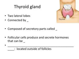
Thyroid Gland Digital Laboratory. It’s best to view this in Slide Show mode, especially for the quizzes. This module will take approximately 30 minutes to complete. After completing this exercise, you should be able to: identify, at the light microscope level, each of the following:

E N D
Thyroid Gland Digital Laboratory It’s best to view this in Slide Show mode, especially for the quizzes. This module will take approximately 30 minutes to complete.
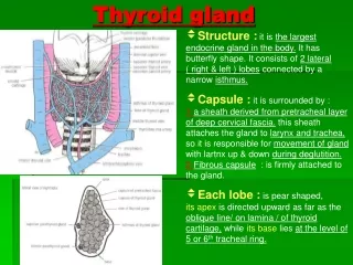
After completing this exercise, you should be able to: • identify, at the light microscope level, each of the following: • Thyroid gland • Follicle • Colloid • Follicular cells • Parafollicular cells (aka C-cells, difficult to identify with routine staining) • identify, at the electron microscope level • Thyroid gland • Follicular cell and all sub-cellular components important for thyroid hormone synthesis • Colloid • Parafollicular cells (C-cells)
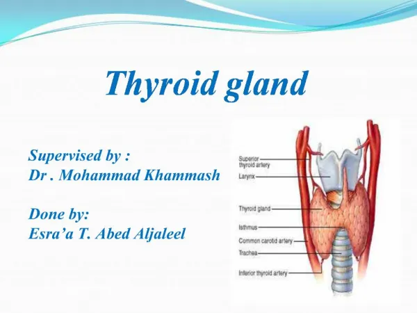
THYROID GLAND DEVELOPMENT These are sagittal views of the developing embryo (A-C) and of the adult (D). The cranial end of the embryo is to the left (neck is flexed). Yellow is the lumen of the digestive tract, in this image showing the future oral cavity and pharynx. Superior surface of the tongue. The thyroid gland develops from the foramen cecum of the tongue. Epithelial cells migrate from the foramen cecum to their final position anterior to the trachea. As they migrate, they are connected to the foramen cecum by the thyroglossal duct, which normally degenerates. Consistent with its epithelial origin, the main thyroid cell type forms a single layer of epithelium that surrounds a “lumen” filled with colloid, and is surrounded by connective tissue.
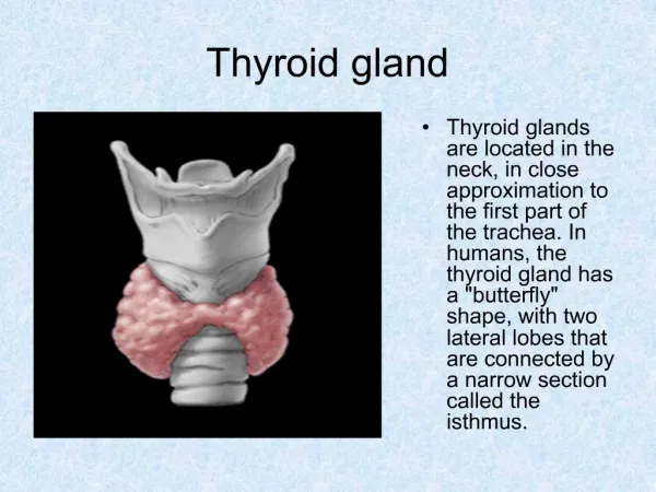
THYROID GLAND A medium-powered view of the thyroid gland shows that it is composed of numerous thyroid follicles (F). Between the follicles is loose connective tissue (CT) that contains numerous blood vessels (BV). Note the follicles are lined by epithelial follicular cells (aka principal cells), and filled with colloid. The clusters of cells indicated by the arrows appear this way because of tangential sectioning through the wall of a follicle.
THYROID GLAND colloid colloid colloid Enlargement of an area similar to that in the rectangle shows more details of the follicles. Although many follicular cells are cuboidal (blue arrows), some are squamous (green arrows). The height of the cells is related to their physiologic activity and thus, in very active glands, follicular cells may be columnar. The follicles are filled with extracellular colloid, which contains a precursor to mature thyroid hormone, called thyroglobulin. Pale cells (black arrow) may be clear cells (C-cells, parafollicular cells), though it is difficult to identify them definitively by H&E. Follicular cells are indeed epithelial, with their apical side facing the colloid, and their basal side facing the connective tissue surrounding the follicles.
THYROID GLAND Video of thyroid gland – SL124 • Link to SL 124 • Be able to identify: • Thyroid gland • Colloid • Follicular cells • (parafollicular cells not definitively identified)
THYROID GLAND Three follicular cells are shown in this electron micrograph of a portion of a thyroid follicle . Like all epithelia, follicular cells sit on a basement membrane (FBL here), which separates these cells from the loose connective tissue surrounding the follicles. The connective tissue in this and other endocrine glands is richly vascularized, as indicated by the capillary lumen (En = endothelial cell of capillary, EBL = basement membrane of endothelial cell). Colloid is found on the apical side of the follicular cells. To ensure separation of colloid from the connective tissue, follicular cells are apically-joined by junctional complexes (JC).
THYROID GLAND Thyroid hormone begins with the production of thyroglobulin, a glycoprotein synthesized in the rER, processed by the Golgi (G), and released into the colloid by exocytosis. Iodide is moved from the blood into the colloid using transport proteins on the basal and apical membranes of the follicular cells. Within follicular cells, iodide is converted to iodine and then, on the microvillus (Mv) membrane, iodine is enzymatically added to tyrosine residues of thyroglobulin. Upon stimulation by thyroid stimulating hormone (TSH), thyroglobulin is brought back into the cell by endocytosis. Endocytic vesicles fuse with lysosomes (L), creating colloidal resorption droplets (CRD). Here, thyroglobulin is degraded, and the resulting iodinated tyrosines are released as thyroid hormones T3 and T4 into the bloodstream.
THYROID GLAND This is from your textbook and may be helpful….
THYROID GLAND - CLEAR CELLS In this micrograph, a parafollicular cell (PC) (aka clear cell or C-cell) is shown surrounded by follicular cells (FC). C-cells typically are smaller than follicular cells, adjacent to the basal lamina, and do not reach the lumen (similar to stem cells in pseudostratified epithelium, though the epithelium of the thyroid is officially simple cuboidal). Interestingly, C-cells are derived from a different source than the foramen cecum. Neural crest cells invade the thyroid during development and give rise to the C-cells. Unlike the follicular cells, which secrete thyroglobulin constitutively into the colloid (C), C-cells store calcitonin in secretory granules, which are often localized on the basal aspect of the cell. They release their product basally into the bloodstream. G=Golgi
The next set of slides is a quiz for this module. You should review the structures covered in this module, and try to visualize each of these in light and electron micrographs. • identify, at the light microscope level, each of the following: • Thyroid gland • Follicle • Colloid • Follicular cells • Parafollicular cells (aka C-cells, difficult to identify with routine staining) • identify, at the electron microscope level • Thyroid gland • Follicular cell and all sub-cellular components important for thyroid hormone synthesis • Colloid • Parafollicular cells (C-cells)
Final quiz Self-check: If this image was taken from the adrenal gland, from which part of that gland could this have been obtained. (advance slide for answers) Adrenal medulla
Final quiz Self-check: This image was taken of the thyroid gland. Identify the cell. Which side of the image is apical? Identify the cytoplasmic organelles important for its activity (advance slide for answers) Lysosomes (or colloid resorption droplets – do not worry about distinguishing the two) Follicular cell RER Golgi apical
Final quiz Self-check: Identify the organ.(advance slide for answers) Adrenal gland
Final quiz Self-check: Where are iodide pumps? (advance slide for answer) Basal side of cell somewhere over here
Final quiz Self-check: Where is thyroglobulin iodinated?(advance slide for answer)
Final quiz Self-check: Where is thyroglobulin synthesized?(advance slide for answers)
Final quiz Self-check: Where is thyroglobulin post-translationallymodified? (advance slide for answer)
Final quiz Self-check: Where is T3 and T4 produced?(advance slide for answers) Hard to say whether these are primary lysosomes or colloid resorption droplets
Final quiz Self-check: Identify the organ. Be specific. (advance slide for answers) Posterior pituitary
Final quiz Self-check: Identify the organ. Be specific. (advance slide for answers) Thyroid gland Please tell me you did not miss this
Final quiz Self-check: If this image was taken from the adrenal gland, from which part of that gland could this have been obtained. Identify 3, 4, and 9. (advance slide for answers) Adrenal cortex 3. Mitochondria with tubular crista 4. Lipid droplets 9. SER
Final quiz Self-check: Identify cells 1 and 2.(advance slide for answers) Follicular cell C-cell