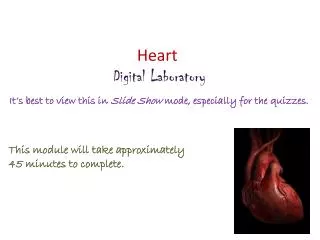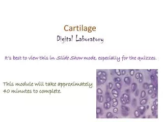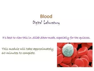
Vessels Digital Laboratory
Vessels Digital Laboratory. It’s best to view this in Slide Show mode, especially for the quizzes. This module will take approximately 40 minutes to complete .

Vessels Digital Laboratory
E N D
Presentation Transcript
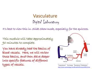
Vessels Digital Laboratory It’s best to view this in Slide Show mode, especially for the quizzes. This module will take approximately 40 minutes to complete. Note that much of this module was presented by necessity earlier in the year, but is placed here again for completeness and reinforcement.
After completing this exercise, you should be able to: • Distinguish, at the light microscope level, each of the following:: • Arteries • Veins • Capillaries • Lymphatic channels
Blood and lymphatic vessels Vessels in the body transport fluids, either blood or lymphatic fluid, which allows for distribution of nutrients, waste products, hormones, etc. throughout the body. There are two components of the vascular system: Cardiovascular system – transports blood, and includes the heart, arteries, capillaries, and veins Lymphatic system – which returns tissue fluid (lymph) from the tissues back to the cardiovascular system
Blood and lymphatic vessels Both of these systems are essentially a set of tubes, with different characteristics. The contents of these tubes, namely blood and lymph, will be covered elsewhere. Here, we focus on the tubes themselves. However, at this time, we will focus on basic characteristics of arteries, capillaries, veins, and lymphatic channels. More details regarding the histology of the cardiovascular system will be presented in the Cardiovascular, Renal, and Pulmonary Block (CRaP).
Blood and lymphatic vessels As mentioned, the details of the different types of arteries and veins will be presented elsewhere. Here, note that vessels have three basic components (from inside to outside): Tunica intima – a simple squamous epithelium, called the endothelium, with underlying loose connective tissue Tunica media – a thicker layer with smooth muscle and elastic fibers Tunica externa(adventitia) – dense connective tissue
Blood and lymphatic vessels In this cross section of a blood vessel, the blood in the lumen is indicated. The double-arrow indicates the extent of the tunica media; you should recognize the smooth muscle tissue that is predominant in this region. The smooth muscle cells are circularly arranged; in other words, the cells lie perpendicular to the long axis of the vessels. The thin portion of the wall “inside” the tunica media is the tunica intima, while the dense irregular connective tissue surrounding the tunica media is the adventitia. blood
Blood and lymphatic vessels In the higher-powered image, it is easier to see the tunica intima (green double-arrow), tunica media (black double-arrow) and adventitia (blue double arrow). Arrows indicate nuclei of endothelial cells that form the inner lining of vessels. blood The border between the intima and media (yellow dotted line) is indeed wavy here; we’ll address this in detail later.
Blood and lymphatic vessels Video of vessel layers – SL85 • Link to SL 085 • Be able to identify: • Blood vessel • Tunica intima • Tunica media • Tunica externa (adventitia)
Blood and lymphatic vessels • In this overview, we will focus on a few key differentiating features of vessels: • The major histological difference between arteries and veins lies in the thickness and muscularity of the tunica media; arteries have a thicker, more muscular tunica media • Capillaries are composed simply of endothelial cells, without a tunica media or adventitia • Lymphatic vessels have an even less-developed tunica media, and the smallest lymphatic vessels have valves
Blood and lymphatic vessels Histologically, differentiating between arteries/arterioles (red arrow) and veins/venules(blue arrow) is best done by comparing vessels of approximately the same size. Arteries have a thicker smooth muscle layer in their wall; therefore, their wall is relatively thicker compared to the size of the vessel itself, with a narrower lumen. In addition, arteries tend to be rounder. Both will typically contain blood cells in their lumen, though during tissue preparation they can be washed away (see vessel toward the left) and become trapped in inappropriate locations. Arterioles are smaller arteries; venules are smaller veins.
Blood and lymphatic vessels Higher-powered view of the same vessels… All blood vessels are lined by a simple squamous epithelium, referred to as an endothelium. Endothelial cells have flattened nuclei (black arrows). The “fatter” nuclei in the wall of the vessel (green arrow), particularly in the artery, belong to smooth muscle cells.
Blood and lymphatic vessels Capillaries are very thin-walled, small vessels; they consist mostly of the endothelial cells. Because of this, they are difficult to find and identify definitively. Possible candidates for capillaries are indicated by the arrows, though the left is likely a small venule, and the right a small arteriole.
Blood and lymphatic vessels A couple more examples of capillaries. In the left image, note the simple squamous endothelial cells that make up the wall, and a diameter that is approximately the size of a red blood cell (possible RBC in upper capillary). To the right is a longitudinal section through a capillary (green arrows).
Blood and lymphatic vessels Lymphatic vessels (green arrows) typically have even thinner walls than veins, although these examples show relatively thick walls. However, small lymphatic vessels in tissues have numerous valves. In addition, they are typically devoid of red blood cells, and are filled with a “colloid” from precipitated lymph fluid as well as white blood cells. Yes, veins have valves too, but not in the smallest veins in the wall of an organ like we are looking at now. artery
Blood and lymphatic vessels Enlarged portion of previous image. If you look VERY closely in the artery, you might see that the pink disks are red blood cells, whereas the pink in the lymph vessels isn’t cellular. In addition, note the much greater proportion of white blood cells in the lymphatic vessels (green arrows) compared to the artery. artery
Blood and lymphatic vessels Video of lung hilus showing blood and lymphatic vessels – SL24 • Link to SL 024 • Be able to identify: • Artery / arteriole • Vein / venule • Capillary • Lymphatic channel
The next set of slides is a quiz for this module. You should review the structures covered in this module, and try to visualize each of these in light and electron micrographs: • Distinguish, at the light microscope level, each of the following: • Arteries • Veins • Capillaries • Lymphatic channels
Final quiz Self-check: Identify the outlined structures. (advance slide for answer) Artery / areteriole
Final quiz Self-check: Identify the outlined structure (advance slide for answer) vein
Final quiz Self-check: Identify the outlined structure (advance slide for answer) Serous gland
Final quiz Self-check: Identify the outlined structure (advance slide for answer) vein
Final quiz Self-check: Identify the structure indicated by the arrows(advance slide for answer) arteriole
Final quiz Self-check: Identify the outlined structure. (advance slide for answer) vein
Final quiz Self-check: Identify the predominant tissue on this slide. (advance slide for answer) Pseudostratified ciliated columnar epithelium (with goblet cells) That sure is one good-looking epithelium; the cilia are fine.
Final quiz Self-check: Identify the outlined structure (advance slide for answer) Lymphatic vessel
Final quiz Self-check: Identify the structure indicated by the arrows (advance slide for answer) Capillary
Final quiz Self-check: Identify the predominant tissue in the outlined region (advance slide for answer) Unilocular adipose
Final quiz Self-check: Identify the outlined structure (advance slide for answer) Lymphatic vessel
Final quiz Self-check: Identify the predominant tissue on this slide (advance slide for answer) Skeletal muscle
Final quiz Self-check: Identify the outlined structures. (advance slide for answer) lymphatic vessel Vein/venule Artery/arteriole
Final quiz Self-check: Identify the outlined structures. (advance slide for answer) vein / venule
Final quiz Self-check: Identify the outlined structure (advance slide for answer) artery
Final quiz Self-check: Identify the structure indicated by the arrows(advance slide for answer) venule
Final quiz Self-check: Identify the outlined structure (advance slide for answer) Artery
Final quiz Self-check: Identify the outlined structure (advance slide for answer) mucous gland (with serous demilune)