Download
1 / 42
430 likes | 936 Views
D.Reshaid Aljurayyan Assit. Professor department of radiology college of medicine King Saud university. Radiology & investigation of hepatobiliary system. Lecture outline:. What is hepatobiliary system HBS? Radiological modalities used in imaging HBS.

E N D
D.Reshaid Aljurayyan Assit. Professor department of radiology college of medicine King Saud university Radiology & investigation of hepatobiliary system
Lecture outline: • What is hepatobiliary system HBS? • Radiological modalities used in imaging HBS. • Advantages and disadvantages of each radiology modality. • Indications of imaging HBS.
What is hepatobiliary system (HBS)? It includes liver, gallbladder and biliary ducts.
What are the Radiological modalities used in imaging HBS ? • X Ray. • Ultrasound. • Computed tomography CT scan. • Magnetic resonance imaging MRI. • Nuclear scan.
What is this? Abdomen x-ray OR Abdomen radiography
X ray was first observed and documented in 1895 by Wilhelm Conrad Roentgen First x ray taken in history
What is X ray? • It is energetic form of electromagnetic and ionozintng radiation that can penetrated solid objects and used to take images of the human body.
X RAY language • Radio-lucent = black • Radio-opaque= white
X RAY Advantages: • Quick and widely available • Cheap • Can be done bedside (portable) Disadvantages: • Use ionizing radiation • Very poor in tissue details
What is this? ULTRASOUND
What is US? • A diagnostic technique in which high-frequency sound waves penetrate the body and produce multiple echo patterns. • Diagnostic Medical applications in use since late 1950’s
Echo pattrens Hyper-echoic = White Hypo-echoic = Light Grey An-echoic = Black
Ultrasound Advantages : • No radiation • Widely available • Relatively cheap • Very good in evaluating abdomen solid organs • Can be done bedside (portable) Disadvantages: • Operator dependent • Very limited in evaluating structures with air ( e.g. bowel) or calcification (e.g. bone)
B- MODE. DUPLEX COLOR DOPPLER
DUPLEX B- MODE
What is this? Computed Tomography CT scan.
What is CT scan? • A CT scan makes use of computer-processed of many X-ray images taken from different angles to produce cross-sectional tomographic images of specific areas of a scanned object. • CT scan can be done with and without intravenous IV contrast. • CT scan is limmited in evaluating gallstones, Why?
What is different between the tow iamges? With IV contrast Without IV contrast
CT language • Hyper-dense = white • Hypo-dense=black to grey
Computed tomography CT scan. Advantages: • Very good in evaluating solid organs • Available more them MRI Disadvantages: • Use ionizing radiation • Less available then x-ray and US • Relatively expansive • Intravenous contrast maybe harmfull
What is this? • Magnetic resonance imaging MRI
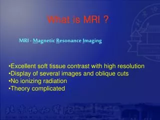
Magnetic resonance imaging MRI • A medical imaging technique used in radiology to form pictures of the anatomy using strong magnetic fields and radio waves. • It has no radiation. • It more complex then CT scan and many different images (or what called sequences) can by taken like T1 and T2 etc.
MRI language • Hyper intense signal = more white • Hypo intense signal = more grey/black
Magnetic resonance imaging MRI Advantages: • Excellent in tissue details • No ionizing radiation Disadvantages: • Expensive • Long scan time • Less available then other modalities • Intravenous contrast is not safe with poor renal function.
Different MRI sequences T2 Diffusion T1
What is this? Nuclear scan
What is nuclear medicine? • Medical specialty involving the application of radioactive substances in the diagnosis and treatment of disease.
Nuclear medicine: Advantages: • Excellent in evaluating oragn function/physiology Disadvantages: • Use ionizing radiation • Not widely available • Very poor in evaluating anatomy