Download
1 / 61
700 likes | 1.22k Views
Lecture 4 Structure and function of nucleic acid. Contents. Composition of nucleic acid Structure and function of DNA Structures and functions of RNA Properties of nucleic acid. Brief history. 1869: isolated DNA from salmon sperm (Friedrich Miescher)

E N D
Contents • Composition of nucleic acid • Structure and function of DNA • Structures and functions of RNA • Properties of nucleic acid
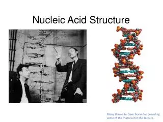
Brief history • 1869: isolated DNA from salmon sperm (Friedrich Miescher) • 1944: proved DNA is genetic materials(Avery et al.) • 1953: discovered DNA double helix(Watson and Crick) • 1968: decoded the genetic codes(Nirenberg) • 1981: invented DNA sequencing method(Gilbert and Sanger) • 1987: launched thehuman genome project • 2001: accomplished the draft map of human genome
Deoxyribonucleic acid, DNA Nucleic acid Ribonucleic acid, RNA
1. The components of DNA and RNA • DNA and RNA are polymers of nucleotide units. • DNA (RNA) consists of 4kinds of ribonucleotide units linked together through covalent bonds. • Each nucleotide unit is composed of • a nitrogenous base • a pentose sugar • a phosphate group
1.1 Bases • Purines : • Adenine (A) • Guanine (G) • Pyrimidines : • Cytosine (C) • Uracil (U) • Thymine (T) DNA: A,G,C,T RNA: A,G,C,U Thymine (T) is a 5-methyluracil (U)
1.2 Ribose (in RNA) and deoxyribose (in DNA) • Ribose and deoxyribose predominantly exist in the cyclic form.
1.3 Nucleosides =ribose/deoxyribose + bases • The bases are covalently attached to the 1’ position of a pentose sugar ring, to form a nucleoside Glycosidic bond 1 R Ribose or 2’-deoxyribose
1.4 Nucleotides = nucleoside + phosphate • A nucleotide is a nucleoside with one or more phosphate groups bound covalently to the 3’-, 5’, or ( in ribonucleotides only) the 2’-position. In the case of 5’-position, up to three phosphates may be attached. Phosphate ester bonds Ribonucleotides (containing ribose) Deoxynucleotides (containing deoxyribose)
phosphate pentose nucleic acid nucleotides nucleosides bases
1.5 Some important nucleotides • dATP, dGTP, dCTP, dUTP • Raw materials for DNA biosynthesis. • ATP, GTP, CTP, UTP • Raw materials for RNA biosynthesis • Energy donor • Important co-enzymes • Cycling nucleotides—cAMP, cGMP • Secondary messengers in hormones action.
AMP ADP ATP Nucleic acid derivatives Multiple phosphate nucleotides • adenosine monophosphate (AMP) • adenosine diphosphate (ADP) • adenosine triphosphate (ATP)
2. Structure and function of DNA 2.1 Primary structure • Definition: the base sequence (or the nucleotide sequence) in polydeoxynucleotide chain. • The smallest DNA in nature is virus DNA. The length of φX174 virus DNA is 5,386 bases (a single chain). • The DNA length of human genome is 3,000,000,000 pair bases.
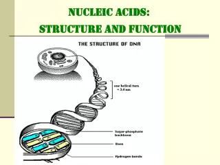
5’end Phosphodiester bond 3’ end: free hydroxyl (-OH) group • 3’,5’ phosphodiester bond link nucleotides • together to form polynucleotide chains
The structure of a DNA chain can be concisely represented • An even more abbreviated notation for this chain is • pApCpGpTpA • pACGTA • The base chain is written in the 5’ →3’ direction
2.2 Secondary structure The secondary structure is defined as the relative spatial position of all the atoms of nucleotide residues.
Secondary structure — DNA double helix structure Francis H.C. Crick • Watson and Crick , 1953 • The genetic material of all organisms except for some viruses. • The foundation of the molecular biology. James D. Watson
The discovery of DNA double helix • Chargaff's Rule (A=T, G=C in DNA) • Franklin, Wilkins: X-ray DiffractionRefined Structure
DNA double helix Essential for replicating DNA and transcribing RNA • Two separate strands • Antiparellel(5’3’ direction) • Base pairing: hydrogen bonding that holds two strands together • Complementary (sequence) 3’ 5’ • Sugar-phosphate backbones (negatively charged): outside • Basepairs (stack one above the other): inside 3’ 5’
4 1 3 2 6 7 5 8 1 9 4 2 3 A:T G:C Base pairing
B form of DNA double helix • Right-handed helix; • The diameter of the double helix:2 nm • The distance between two base pairs: 0.34 nm; • Each turn of the helix involves 10 bases pairs, 3.4 nm. • Stable configuration can be maintained by hydrogen bond and base stacking force (hydrophobic interaction).
Groove binding • Small molecules like drugs bind in the minor groove, whereas particular protein motifs can interact with the major grooves.
Watson, Crick, and Wilkins shared the Nobel Prize in medicine or physiology in 1962 for this brilliant accomplishment. • The discovery of the DNA double helix revolutionized biology: it led the way to an understanding of gene function in molecular terms (their work is recognized to mark the beginning of molecular biology).
Conformational variation in double-helical structure • B-DNA • A-DNA • Z-DNA
B-form: the duplex structure proposed by Watson and Crick is referred as the B-form DNA. • It is the standard structure for DNA molecules. • A-form: at low humidity the DNA molecule will take the A-form: • The A-form helix is wider and shorter, with a shorter more compact helical structure, than the B-form helix. • Z-form: the Z-form DNA is adopted by short oligonucleotides. • It is a left-handed double helix in which backbone phosphates zigzag.
2.3 Tertiary structure : • Supercoils: double-stranded circular DNA form supercoils if the strands are underwound (negatively supercoiled) or overwound (positively supercoiled). Increasing degree of supercoiling Relaxed supercoiled
If the strands are overwound, form positively supercoiled; • If the strands are underwound, form negatively supercoiled.
The DNA in a prokaryotic cell is a supercoil. • Supercoiling makes the DNA molecule more compact thus important for its packaging in cells.
2.4 Eukaryotic DNA • DNA in eukaryotic cells ishighly packed. • DNA appears in a highly ordered form called chromosomes during metaphase, whereas shows a relatively loose form of chromatin in other phases. • The basic unit ofchromatin is nucleosome. • Nucleosomes are composed of DNA and histone proteins.
Nucleosome • The chromosomal DNA is complexed with five typesof histone. • H1, H2A, H2B, H3 and H4. • Histons are very basic proteins, rich in Arginine and Lysine. • Nucleosomes: regular association of DNA with histones to form a structure effectively compacting DNA. ”beads”
Beads on a string • 146 bp of negatively supercoiled DNA winds 1 ¾ turns arounda histone octomer. • H1 histone binds to the DNA spacer.
The importance of packing of DNA into chromosomes • Chromosome is a compact form of the DNA that readily fits inside the cell • To protect DNA from damage • DNA in a chromosome can be transmitted efficiently to both daughter cells during cell division • Chromosome confers an overall organization to each molecule of DNA, which facilitates gene expression as well as recombination.
2.5 Functions of DNA • The carrier of genetic information. • The template strand involved in replication and transcription. Gene: the minimum functional unit in DNA Genome: the total genes in aliving cell or living beings.
3. Structures and functions of RNA Conformational variability of RNA is important for the much more diverse roles of RNA in the cell, when compared to DNA. Types : • mRNA: messenger RNA, the carrier of genetic information from DNA to translate into protein • tRNA: transfer RNA , to transport amino acid to ribosomes to synthesize protein • rRNA: ribosomal RNA, the components of ribosomes • hnRNA: Heterogeneous nuclear RNA • snRNA: small nuclear RNA
RNA structure • RNA molecules are largely single-stranded but there are double-stranded regions.
3.1 Messenger RNA( mRNA) • Function: the carrier of genetic information from DNA for the synthesis of protein. • Comprises only about 5% of the RNA in the cell. • Composition: vary considerably in size (500-6000 bases in E. coli)
Eukaryotic mRNA Structure • Capping: linkage of 7-methylguanosine to the 5’ terminal residue. (2) Tailing: attachment of an adennylate polymer (poly A, 20~250 nucleotides) at the 3’ terminal.
3.2 Transfer RNA (tRNA) • They make up 15% of the RNA in the cell. • Function:Transport amino acids to ribosomes for assembly into proteins. • There are at least 20 types of tRNA in one cell. • Primary Structure : • 74~95 bases, the smallest of the three major RNA. • Modified bases: pseudouridine (ψ) methylguanosine dihydrouridine (D) • The sequence CCA at the 3’ terminus
Secondary structure: cloverleaf • Four loops and four arms • Amino acid arm (7bp): to bide amino acid • D loop(8-14bp) and D arm(3-4bp): • Anticoden loop(5bp) and arm(7bp): to recognize amino acid coden on the mRNA. • TψC loop(7bp) and arm(5bp) • Variable loop(4-5bp or 13-21bp)
3.3 Ribosomal RNA (rRNA) • Components of ribosomes. • They make up 80% of the RNA in the cell. * The species of rRNA • Eukaryotes • 5S rRNA • 28S rRNA • 18S rRNA • 5.8S rRNA • Prokaryotes • 5S rRNA • 23S rRNA • 16S rRNA • S represents Svedberg units, they represent measures of sedimentation rate.
The proposed secondary structure for E.coli 16S rRNA
Ribosomes • Ribosomesare cytoplasmic structures that synthesize protein, composed of RNA (2/3) and protein (1/3). • The ribosomes of prokaryotes and eukaryotes are similar in shape and function. The difference between them is the size and chemical composition.