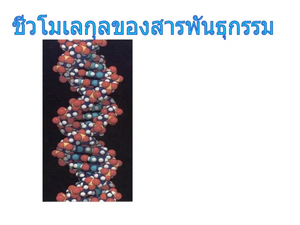
Biochemistry for Pharmacy Students NUCLEIC ACID STRUCTURE & FUNCTION
1.12k likes | 1.48k Views
Biochemistry for Pharmacy Students NUCLEIC ACID STRUCTURE & FUNCTION. Pál Bauer Dept. of Medical Biochemistry Rm. 4 515, EOK Email: bauerpal@hotmail.com. OBJECTIVES. To learn the structures and properties of nucleotides, the building blocks of nucleic acids

Biochemistry for Pharmacy Students NUCLEIC ACID STRUCTURE & FUNCTION
E N D
Presentation Transcript
Biochemistry for Pharmacy StudentsNUCLEIC ACID STRUCTURE & FUNCTION Pál Bauer Dept. of Medical Biochemistry Rm. 4515, EOK Email: bauerpal@hotmail.com
OBJECTIVES • To learn the structures and properties of nucleotides, the building blocks of nucleic acids • To describe how the structure of DNA relates to functions of the genetic machinery. • To explain how DNA is synthesized. • To describe how mutations in DNA can lead to genetic diseases. • To explain how recombinant DNA technology can be applied to the diagnosis and therapy of human genetic diseases (discussion sessions).
HISTORICAL PERSPECTIVE The beginnings of nucleic acid biochemistry & genetics occurred in the 1860s: • Frederic Miescher – Swiss biochemist who • discovered nucleic acids. • Gregor Mendel – Austrian monk who founded the • science of genetics. Nucleic acid structure & metabolism is directly relevant to cancer, gout, many genetically inherited diseases, AIDS and other infectious diseases.
Gregor Mendel studied garden peas
Sugars and phosphates • Ribose in RNA • Deoxyribose in DNA • Partial hydrolysis products • Nucleotides (contains phosphates) • Nucleosides (no phosphates)
Base composition of double stranded DNA • [pyrimidines] = [purines] • Content: A = T, G = C • DNA base compositions are the same for different tissues of the same organism.
Crick Watson
A Reminder of the Differences between Eukaryotes and Prokaryotes Animal Cell (eukaryote) E. coli Bacterial Cell (prokaryote) Plant Cell (eukaryote) Fig. 2-7
Model of the Nuclear Envelope Artwork by Don Guzy
5’ 3’ • The Watson-Crick Structure • Right-handed helices • Anti-parallel • Base-pairing • Open structure accessible to water • Stacking forces between planar paired bases give a rigid structure 5’ 3’
Denaturation of DNA By pH, heat, solvents, urea, amides • Helix-coil transition • Hyperchromic effect • Melting temperature
Denaturation of double-stranded DNA
DNA IS THE GENETIC MATERIAL IN CELLS Indirect evidence • High DNA content of chromosomes • 260 nm is a very mutagenic wavelength; bases maximally absorb light energy at 260 nm • Constancy of [DNA] / cell (germ cells) and 2x [DNA] / cell (somatic cells)
Direct Evidence a. Transformation of bacteria with DNA Requires both DNA functions: replication and expression b. Transfection with viral nucleic acids c. In vitro expression of DNA (from bacteriophage T4) d. Synthesis of active DNA in vitro
DNA CONTENT OF SOME CELLS AND VIRUSES Haploid size of genome, Source of DNA base pairs____ Viruses SV405 x 103 (5 kb) Papilloma (wart)8 x 10³ Adenoviruses2.1 x 104 Herpesviruses1.56 x 105 Poxviruses2.4 x 105 Cells Escherichia coli4.5 x 106 (4,500 kb) Yeast1.3 x 107 Drosophila1.6 x 108 Human 3.2 x 10⁹(3.2x10⁶kb) Frogs6 x 109 (6 x 10⁶ kb) OnionPlant18 x 109 Fern Plant160 x 109 Animal mitochondria 1.5 x 10⁴ (15 kb) Plant chloroplast 1 x 10⁵
The total length of the circular E. coli chromosome is 1.7 mm, whereas the length of an E. coli cell is 2 μm. partially lyzed E. coli cell
Human haploid genome 22 autosomal chromosomes (chromosomes 1-22) plus X and Y chromosomes 3.2 x 10⁹ total bp
The double- stranded DNA is wrapped around the outside of each nucleosome twice. The nucleosomes are regularly spaced along the DNA. electron micrograph of chromatin
electron micrograph of supercoiled nucleosomes in chromatin.
A typical phone cord is coiled like a DNA helix, and the coiled cord can itself coil in in a supercoil. If twisted tight enough, the supercoils will themselves form an even higher order of supercoiling. Double-stranded DNA helices also form supercoils of supercoils.
Two drawings depicting the different levels of DNA supercoiling that provide DNA compaction in a eukaryotic chromosome. The levels take the form of coils upon coils. nucleosomes histones
DNA replication 3’ 5’ 5’ 3’ 3’ 5’ 5’ 5’ 3’ 5’ 3’ 3’
5’ 3’ 5’
All DNA polymerases catalyze elongation of the primer strand in a 5’ to 3’ direction, copying the template strand in a 3’ to 5’ direction. This means that the “leading strand” can be synthesized continuously, but the “lagging strand” must be synthesized discontinuously. A problem is that DNA polymerases must have a primer strand with a 3’ OH from which to begin DNA synthesis. So, where do these primers come from when a DNA polymerase synthesizes new Okazaki fragments in the lagging strand?
3’ 5’ 5’ 3’ 3’ 5’ 5’ 3’ Replication does not usually begin at the end of a DNA molecule; it begins in the middle of the DNA. (DNA pol I, II and III) DNA
12.4 Schematic model of the proofreading function of DNA polymerase Figure 12-21
An example of error correction by the 3’ to 5’ exonuclease (proofreading) activity of DNA polymerase I. A mismatched base pair (a C-A mismatch) impedes the movement of DNA polymerase I to the next site. Sliding back- ward, the enzyme removes its mistake with the 3’ to 5’ exo- nuclease activity & then resumes its 5’ to 3’ polymerase activity.
c. DNA Polymerase II • Mutants in the E. coli gene for this DNA • polymerase are not lethal. Also is a repair • enzyme. • Requires duplex DNA template and primer. • Utilizes a template strand and elongates a primer • strand, similar to DNA polymerase I. d. DNA Polymerase III (pol Cgene) Mutants are ts (temperature sensitive), i.e., conditionally lethal.
The 3-D structure of E. coli DNA polymerase I. The active site for the polymerase activity and the 3’ to 5’ exonucelase activity is deep in the crevice at the far end of the bound DNA. The template strand is dark blue.
a. Illustration of the 5’ to 3’ exonuclease activity of E. coli DNA polymerase I (sometimes called a “nick translation” activity). An RNA or DNA strand paired with a DNA template strand is simultaneously degraded by the 5’ to 3’ exonuclease activity & is replaced by the polymerase activity of the same enzyme.
b. The origin of replication – the oriC locus Accessory proteins dna B gene product Probably this protein is membrane-associated and recognizes the initiation sequence on DNA. c. RNA primers DNA polymerases require a preformed primer; RNA polymerases do not. d. Details of the events: 1. Accessory protein binds (dna B protein) 2. "Primase," an RNA polymerase (dna G) It does not require a primer; forms a short piece of RNA complementary to the DNA template strand. 3. DNA binding proteins
cell membrane cell membrane
5’ 5’ 5’ 3’ 3’ 3’ 5’ 5’