Download
1 / 42
420 likes | 580 Views
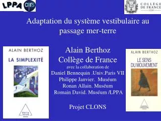
Adaptation du système vestibulaire au passage mer-terre Alain Berthoz Collège de France avec la collaboration de Daniel Bennequin .Univ.Paris VII Philippe Janvier. Muséum Ronan Allain. Muséum Romain David. Muséum /LPPA Projet CLONS. I . Le système vestibulaire, un référentiel mobile.

E N D
Adaptation du système vestibulaire au passage mer-terreAlain BerthozCollège de Franceavec la collaboration de Daniel Bennequin .Univ.Paris VIIPhilippe Janvier. MuséumRonan Allain. Muséum Romain David. Muséum /LPPAProjet CLONS
I . Le système vestibulaire, un référentiel mobile
Une simplification fondamentale: séparation des rotations et des translations (From B. Hess and D.Angelaki 1999)
Saccule et utricule Jaeger-Haslwanter 2002,2004 Moravec, Xue, Peterson, 2006 Wilson, Melville Jones, Curthoys,…
Cyclostomes (living jawless vertebrates) www.uvm.edu Courtesy P. Janvier Morphology & physiology Gene sequence data
Osteostracans: jawless vertebrates with only two vertical semicircular canals Canal to lateral « fields » Lateral field nerves Marginal artery VII VII V2-3 Nasal capsule IX Marginal vein IX Pineal IX Brachial vein X1 Brachial artery Brachial plexus Endolymphatic duct Labyrinth Norselaspis glacialis , Early Devonian (400Myr, Spitsbergen. Dorsal view
Gnathostomes (jawed vertebrates) Morphology & physiology Gene sequence data
Le canal horizontal apparait …………………….
«Back evolutionary phenotypes», «atavism», knock-outs, and «ancestral morphologies» Otx1-/- Acampora et al. Nature Gen. 1996 Mazan et al. Evol. Devel. 2000 Otx1+/+
Otx1 gene-controlled morphogenesis of the horizontal semicircular canal and the origin of the gnathostome characteristics The horizontal semicircular canal of the inner ear is a unique feature of gnathostomes and is predated by the two vertical semicircular canals, A strong correlation is observed in extant vertebrates between the distribution of the horizontal semicircular canal and the presence of an Otx1 ortholog expressed in the inner ear. Some of the anatomic characteristics of gnathostomes have appeared quite suddenly and almost simultaneously in vertebrate evolution, possibly as a consequence of gene functional diversifications following duplications of an ancestral chordate gene. Sylvie Mazan, Danielle Jaillard, Blandine Baratte, and Philippe Janvier EVOLUTION & DEVELOPMENT 2:4, 186–193 (2000)
Evolution of the perilymphatic duct system and pars inferior of the vertebrate inner ear. • The amphibian inner ear has a simple perilymphatic duct (pd) that begins with the perilymphatic sac (ps) • The elastic perilymphatic duct approximates the extension of the sacculus that contains the basilar papilla and transmits pressure changes to this organ. • B. In the reptile inner ear the perilymphatic duct is more elongated and intimately associated with the elongated lagena.Perilymphatic motion caused by ambient vibration and/or pressure changes is transmitted to an organ specialized to sense these stimuli. • C. In birds, crocodylids, and mammals the perilymphatic duct becomes more elongated and, in mammals, differentiates into scalavestibuli (sv) and scala tympani (st). These envelop the cochlear duct, containing the auditory sensory organ of Corti. Evolutionary Changes in the Cochlea and Labyrinth: Solving the Problem of Sound Transmission to the Balance Organs of the Inner Ear JOHN CAREY* AND NIVEE AMIN Department of Otolaryngology-Head and Neck Surgery, Johns Hopkins University School of Medicine, Baltimore, Maryland
Evolutionary Changes in the Cochlea and Labyrinth: Solving the Problem of Sound Transmission to the Balance Organs of the Inner Ear JOHN CAREY* AND NIVEE AMIN Department of Otolaryngology-Head and Neck Surgery, Johns Hopkins University School of Medicine, Baltimore, Maryland Fig. 1. Sensory transduction by vestibular hair cells. A: At rest, there is some baseline release of excitatory neurotransmitter from the hair cell onto the underlying afferent neuron ending. B: Hair cells are depolar ized when the stereocilia are deflected in the “on” direction (toward the kinocilium in black). C: This occurs because the stretched tip links mechanically open cationic channels in the stereocilia membranes. The influx of potassium ions raises the hair cell’s membrane potential. D: The increased membrane potential activates voltage-sensitive calcium channels in the basolateral membrane of the cell. Synaptic release of excitatory neurotransmitter increases, and receptors in the postsynaptic (psd) density on the afferent increase its membrane potential, which in turn increases afferent firing rate.
Le saccule et l’utricule Les amniotes (Haeckel 1866) sont des vertébrs tétrapodes qui possèdent un amnios ou sac amniotique, protégeant l'embryon ou le foetus. L'embryon, protégé par un œuf à coquille dure ou par l'utérus maternel, se développe en milieu aqueux à l'intérieur de l'amnios. Les amniotes contiennent les sauropsides (dont les oiseaux) et les synapsides (dont les mammifères). Première remarque : chez tous ceux qui sont sortis de l’eau, y compris ceux qui ne sont pas des amniotes (comme les amphibiens), la macula du saccule s’est élargie et s’est remplie de cellules sensorielles.
Teleosts Utriculi Sacculus and Lagenae ( Disparait)
Living jawed vertebrates Sharks, rays and Ratfish (chondrichthyans) Ray-finned fishes (actinopterygians) Coelacanths actinistians Latimeria chalumnae Un intermédiaire Intéressant: le Lungfish Lungfishes (dipnoans) LepidosirenParadoxa-3 Four-legged vertebrates (tetrapods) « Lobe-finned fishes » (sarcopterygians) Courtesy P. Janvier
The Lungfish La striola dans les deux organes possède une des formes “tétrapode”: celle des mammifères!
Frog’s utricle manifold Lewis-Li, 1975, Baird, 1992; Rohregger, Straka, Dieringer, 2002, 2004
L’apparition d’un deuxième système de cellules sensorielles
Deuxième remarque : Chez les amniotes, il est apparu un second système, celui qui est dit de type I, correspondant à des afférents phasiques et irréguliers, à des conductances de courants particulières (par exemple un potassium activé au potentiel de repos), et, pour les otolithes, à une disposition concentrée autour de la striola ou à partir du centre de la crista. Les cellules afférentes de type I, se terminant uniquement avec des calices, irrégulières, phasiques et à faible gain, et une sensibilité plus grande à fréquence plus élevée .(Goldberg, Minor, par exemple 15Hz versus 4Hz pour la crista d’un mammifère)
Pigeon Utricle Saccule
. Chez les modernes téléostes l’utricule est nécessaire pour maintenir l’équilibre vertical, pas le saccule ni la lagena. L’audition des poissons est assez restreinte (en-dessous de 1000Hz), elle met à contribution, en dehors de la ligne latérale, le saccule, la lagena, la macula neglecta, etc. Chez tous les amniotes s’est développé une oreille externe, et du coup l’oreille interne s’est transformée pour transmettre une information auditive .
Les relations entre les capteurs vestibulaires et le système oculomoteur et céphalomoteur.
Les relation entre capteurs vestibulaires et muscles des yeux est maintenue par la connectivité neuronale Courtesy H. Straka et R. Baker (2009)
II . Bases neurales de la capacité de nager et marcher
From swimming to walking: a single basic network for two different behaviors The swimming pattern is generated when the network receives tonic excitation with local intensity gradients near the neck and girdle regions. In the walking pattern, the network receives (in addition to a tonic excitation at the girdles) a phasic drive which is out of phase in the neck and tail regions in relation to the middle part of the body. Tiaza Bem, Jean-Marie Cabelguen, Orian Ekeberg, Sten Grillner Biol. Cybern. 88, 79–90 (2003)
The lamprey locomotor system (Courtesy Sten Grillner)
From Swimming to Walking with a Salamander Robot Driven by a Spinal Cord Model A primitive neural circuit for swimming can be extended by phylogenetically more recent limb oscillatory centers to explain the ability of salamanders to switch between swimming and walking. Auke Jan Ijspeert, Alessandro Crespi, Dimitri Ryczko, Jean-Marie Cabelguen Science 315,1416 (2007);
From Swimming to Walking with a Salamander Robot Driven by a Spinal Cord Model Walking Swimming . Switching from walking to swimming; activity of the CPG model when the drive signal is progressively increased. (A) xi signals from the left body CPG oscillators (oscillators on the right side are exactly in antiphase). Auke Jan Ijspeert, Alessandro Crespi, Dimitri Ryczko, Jean-Marie Cabelguen Science 315,1416 (2007);
Ancestry of motor innervation to pectoral fin and forelimb We present anatomical and embryological evidence that shows pectoral motoneurons also originate in the hindbrain among both basal and derived ray-finned fishes. This conserved pattern for a hindbrain/spinal motor column was likely an adaptation for pectoral control of balance and head orientation in primitive fishes, including those that gave rise to tetrapods. . We propose that Hox genes provided a mechanism for pectoral motoneurons to be evolutionarily labile in position, allowing eventual decoupling from the hindbrain much like their target appendage gained independence from the head. Leung-Hang Ma1,2, Edwin Gilland2,3, Andrew H. Bass2,4 and Robert Baker1,2 Nature (in Press )2010
Leung-Hang Ma Edwin Gilland Andrew H. Bass and Robert Baker Nature (in Press )2010
Hypothèse: l’apparition d’un cou dont l’architecture permet la création d’un plan privilégié: celui du canal horizontal Chat Vidal, Graf, Berthoz
III L’évolution du système vestibulaire jusqu’à l’homme D’un contrôleur de posture à la relation avec autrui!!!!!!!!!!!!!
La création d’un référentiel par l’utilisation de la gravité )
La création d’un plan privilégié horizontal Permet la spécialisation d’un système neuronal de contrôle des mouvements horizontaux D’où rapidité, précision, anticipation…
Simplification : la stabilisation de la tête ce que Marey n’avait pas vu!! HYPOTHESE: La marche est contrôlée à partir de la tête stabilisée grâce au système vestibulaire et au regard. ( Cf Les référentiels mobiles de Elie Cartan) Pozzo, T. Berthoz, A. Lefort L. Normal subjects Exp Brain Res. 82 (1990) 97-106). ¨Pozzo, T. Berthoz A. et al. ; Vestibular patients Exp. Brain Res. 85 (1991) 1-10
Microraptor, the four-winged bird Microraptor Zhang & Zhou 2004 Lower Cretaceous (100Myr) China
L’évolution du système vestibulaire: les oiseaux sont des dinosaures! • R. David ,Al.Berthoz , P. Janvier , R.Allain , • D. Bennequin, J. Droulez (2010) Hypothèse : le système vestibulaire permet le vol en 3D
Rôle du système vestibulaire dans I’intégration multisensorielle, dans la construction du soi, et l’échange avec autrui p < 0.05 corrigé …. Cortex Vestibulaire primaire W. Penfield. The excitable cortex in conscious man Cortex vestibulaire périsylvien P. Kahane, D. Hoffmann, L. Minotti,A.Berthoz Ann Neurol 2003;54:615 –624 Stimulating illusory own-body perceptions O.Blanke, S. Otigue, T. Landis, M. Seeck Nature-vol 419-19 September 2002 Lobel,, et al. J. Neurophysiol. 1996 PIVC Reading the mind from eye gazeA J. Calder, A D. Lawrence, J Keane, Se K. Scott, A M. Owen, I C, Andrew W. YoungNeuropsychologia 40 2002) 1129–38 Dual streams of auditory afferents target L. M. Romanski, B. Tian, J. Fritz, M. Mishkin, P. S. Goldman-Rakic & J. P. Rauschecker nature neuroscience • 2 12 • 1999 M.Zilbovicius et al Allisonet al Trends in Cognitive Sciences 4-7 pp.267-278 2000
Changes in the inner ear of craniates form a morphological series of alterations, starting with the least complicated ear to be found in extant craniates, that of hagfishes. Derived patterns in all crown groups of vertebrates are characterized by increasingly segregated and more specialized sensory epithelia (fig. 1). For example, mammals have evolved a long, coiled cochlea, which enables them to perceive sound over a wider frequency range than other vertebrates [Lewis et al., 1985]. Amphibians are unique in their evolution of a distinct sensor dedicated to sound perception: the amphibian papilla [de Burlet, 1934] derived from the neglected papilla [Fritzsch and Wake, 1988]. Jawed vertebrates have evolved a third semicircular canal and an associated crista organ, which allows them to detect horizontal angular acceleration selectively [Lewis et al., 1985;Fritzsch et al., 2001]. Lampreys and hagfish must compute this information based on stimulation of one or both vertical canals. Ear evolution can thus be characterized by progressive segregation and specialization of sensory epithelia embedded in an ever more complex three dimensional network of tubes and recesses, within which is embedded the more ancient and evolutionarily conserved mechanosensory module. Keeping Sensory Cells and Evolving Neurons to Connect Them to the Brain: Molecular Conservation and Novelties in Vertebrate Ear Development K.W. Beisel and B. Fritzsch Creighton University,