Download
1 / 67
680 likes | 822 Views
Multiphoton Excited Fluorescence Microscopy: Principles, Instrumentation and Some Applications. Outline MPE Photophysics 2) Instrumentation 3) Some Applications of MPE Fluorescence 4) MPE Photodamage Issues 5) 1,2,3 Photon Resolution. Interaction of Light with Matter.

E N D
Multiphoton Excited Fluorescence Microscopy:Principles, Instrumentation and Some Applications
Outline • MPE Photophysics • 2) Instrumentation • 3) Some Applications of MPE Fluorescence • 4) MPE Photodamage Issues • 5) 1,2,3 Photon Resolution
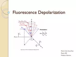
Interaction of Light with Matter P = induced polarization, (n) = nth order non-linear susceptibility E = electric field (3) << (2)<< (1)(5-7 orders of magnitude per term) Linear Processes · Simple Absorption/Reflection · Rayleigh Scattering Third Order Processes · Multi-Photon Absorption* · Stimulated Raman Scattering · Optical Kerr Effect · White Light Generation Second Order Processes · Second Harmonic Generation* · Sum-Frequency Generation
One and two photon absorption physics Goeppart-Mayer, ~1936 Simultaneous absorption Virtual State: Very short lifetime ~10-17 s Requires high power: Absorption only In focal plane e.g. fluorescein Greatly Reduces out of plane bleaching
2-photon excitation of fluorescein: 3D confinement Absorption, Fluorescence only in middle at focal point Compare 1 and 2-p Absorption 1-p excites throughout
Advantages of Non-linear Optical Excitation in Imaging · Intrinsic 3-Dimensionality (no pinhole) · Little Near Infrared and IR Absorption of Biomolecules Greatly Minimizes Out-of-Plane Photo-Bleaching/Damage · Comparable Lateral (X,Y) and Axial (Z) Resolution to confocal · Large Depth of Penetration (scattering decreases in NIR)* 5-10 fold · Enhanced Contrast and Sensitivity* (non-descanned detection) * enabling aspects for tissue imaging: brain, connective tissue, muscle
One and 2-photon absorption characteristics One Photon2 photon d (10-50 cm4s) e (50,000) Absorption Coefficient units 10-50 cm4s= 1 GM (Goppert-Mayer) s (10-16 cm2) p P2 (gives rise to sectioning) Power (photon) dependence Laser Temporal dependence (virtual state) 1/t none Absorption probability p2 s p d /t Cannot use cw lasers (Ar+)
2-Photon Absorption Probability Assume 10 GM cross section (fluorescein) 100 femtosecond pulse 1.4 NA 800 nm 80 MHz Saturation (na≈1) occurs ~50 mW average power Can propagate same form for three-photon absorption: Need at least 10 fold higher average power Do most dyes have good enough two-photon cross sections for imaging?
One photon absorption simply measured in UV-VIS Spectrometer, Beer’s Law A= εcl need new setup for two-photon absorption: need focused light Two-photon cross section measurement Epi geometry Measure Fluor. Measure wavelength Measure pulse width Measure power Control power Xu and Webb, 1996 Measure by fluorescence intensity, need quantum yield (same as 1 photon)
Two-photon spectrum of rhodamine B: Discrete points, not continuous like UV-VIS for 1-photon Max 820 nm not 1050 nm Cross section GM Near-Infrared, rather than visible used for 1-p
Power Dependence to determine photon number: log-log plots Fluorescein and rhodamine Right slope of 2 at All wavelengths: 2-photon process Xu and Webb, 1996
Verify emission spectrum Same emission spectrum for 1-p, 2-p excitation Should be, from Quantum Mechanics Relaxation is independent of Mode of excitation Same emission spectrum For different 2-p wavelengths: 750 and 800 nm Just like 1-photon emission Xu and Webb, 1996
Pulse Width Dependence Slope of 1 Correct pulse width dependence Xu and Webb, 1996
strong medium Rhodamine (best one) fluorescein • Good 2-p properties • Big conjugation • Donor/Acceptor Pair • (push-pull) • Heteroatom substitution Weak UV absorb Small conj Indo-1 2-P δ Rhodamine 5x>fluorescein because D/A pair Also relaxes selection rules over fluorescein
Conclusion: most dyes used for confocal work for Two-photon excitation (some better than others)
Some Generalities about multi-photon absorption • Emission spectrum is the same as 1-p • Emission quantum yield is the same • Fluorescence lifetime is the same • Spectral positions nominally scale for the same transition: • 2-p is twice 1-p wavelength for • 5) Selection rules are often different, especially for xanthenes • (fluorescein, rhodamine, and derivatives, (calcium green, fluos)) Nominally forbidden in 2-p Nominally forbidden in 1-p: missing in fluorescein Allowed and stronger in 2-p
1 and 2-photon bands Reverse of 1-photon For all xanthenes: Fluorescein, rhodamines All max ~830 nm Not ~1000 nm
Fluorescein, Rhodamine Similar band structure Fluorescein Rhodamine B Cross section GM Cross section GM strong medium Xu and Webb, 1996
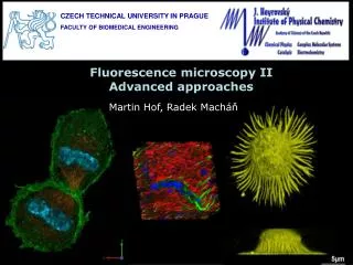
Multi-color imaging in tissue culture cells: Nucleus (blue),mitochondria (red),actin (green) Simultaneous imaging not possible in one photon absorption : Different transitions, need multiple lasers: 390, 490, 540 nm Image all 3 simultaneously via 2-photon with ti:sapphire laser 780 nm: different selection rules for MPE So et al
Confocal detection must be descanned through pinhole to achieve axial discrimination, eliminate out of focus light Very inefficient Optical budget Limited depth of Penetration ~30 microns (based on one scattering Length in most tissues)
2-photon does not Need pinhole at all Or descanning to Eliminate out of Focus light (intrinsic sectioning) Ti:sapphire 100 femtosecond 80 MHZ Open pinhole, Not necessary But still Confocal, lossy Non-decanned Detection greatly Increases the Sensitivity 3-5 fold Very significant Fewer optics No pinhole, Detect scattered light
“The Campagnola” Very simple beampath Relative to confocal
Lasers for multiphoton excitation Typical confocals use fixed laser lines, Can be limiting for multi-color imaging
Lasers for confocal microscopy * * * All cw: no peak power None useful for multiphoton excitation Not tunable or red enough to match 2-photon bands
Tunable Lasers Gain Medium with broad emission spectrum gives tunability to excite any fluorophore Still monochromatic when lasing Organic Dyes (e.g. rhodamines, styryls), titanium sapphire) Ti:sapphire is almost universally used for 2-photon excitation Ti: Al2O3
Titanium Sapphire Ti3+: Al2O3 E 3/2 excited state 2T2 ground state Spin forbidden: long emission lifetime 300 µs ε=14000, strong for spin-forbidden transition Huge Stokes shift, Broad emission spectrum Revolutionized laser industry in ability to make short pulses
Modelocked ti:sapphire laser for 2-photon microscope 2-photon would not have taken off without this laser 100 fs, 80 MHz Tunable 700-1000 nm Pumped with 532 nm
Tuning Range and Power of Ti:Sapphire Longer wavelengths Less damaging 900 nm is often good Compromise between Power and viability Gets dodgy after 900 Except with big laser, Alignment critical Excite essentially every dye, Fluorescent protein With this wavelength range 100 femtosecond pulses 10 nm FWHM bandwidth
Tissue Imaging: MPM enabling Depth and sensitivity
1-p 2-p Contrast stretched 2-photon depth much improved: Reduced scattering 1047 vs 532 nm 2-p descanned here White, Biophys J, 1998
Non-descanned (direct) detection provides greater sensitivity Photons are Scattered, miss pinhole Still fairly Confocal MFP~20-50 microns X-Z projection Sensitivity (best signals) Confocal (1-p)<2-p descanned< 2-p direct 2-p direct collects ballistic and scattered photons White, Biophys J, 1998
Non-descanned (direct) detection provides greater sensitivity Important at increasing depth X-Z direct White, Biophys J, 1998 Make or break experiment with Highly scattering tissue
2-photon imaging of retina (salamander) Fluorescein labeled X-Z projection Too thick for 1-p Good contrast throughout Denk, PNAS, 1999
Depth capability using 2-photon absorption Svoboda, Neuron, 2006, vol 50, 823 (review of 2-P for neuroscience)
Autofluorescence of endogenous species in tissues Need multi-photon excitation, non-descanned detection For enough sensitivity: small cross sections and quantum yields
Autofluorescence in Tumors Mitochondria: NADH, Flavins NAD not fluorescent NADH emission to Monitor respiration NADH good diagnostic Of cell metabolism Small cross section Quantum yield ~10% Small delta ~0.1 GM High concentration Need non-descanned Detection to be viable
Imaging Muscle (NADH) With TPE Fluorescence Low cross section but High concentration Balaban et al
2-photon Tissue Imaging (Mouse Ear) keratinocytes Basal cells Collagen/elastin fibers cartilage So et al Ann. Rev. BME 2000 Autofluorescence good for many layers
Human Skin Two-photon imaging Strata corneum Keratinocytes Dermal layer (elastin, collagen) fibers So et al Ann. Rev. BME 2000 More versatile than dyes (but weaker) MPM enabling, very weak in confocal
Endogenous only way for in vivo clinical Applications. Cannot use dyes (toxicity) cannot penetrate tissues or GFP expressions Trend is multimodal: fluorescence + scattering fluorescence + CT fluorescence + PET Needs multiphoton for depth of penetration and Sensitivity due to weak signals
Multiphoton bleaching Need 3D treatment, both radial, axial PSF
2-photon FRAP in cells, solution Calcein in RBL cells Calcein In solution Webb et al
2-P photobleaching and fluorescence recovery In starfish oocyte 10 kD dye- Dextran Cross nuclear envelope 70 kD does not cross Line scan bleach, page scan recovery Better Cell viability than 1-p due to confinement
2-photon uncaging glutamate Fluo-5 calcium sensitive Alexa Ca insensitive Need 2-p localization For this Svoboda, Neuron, 2006
2-photon photoactivation of GFP Uncaging cross sections very small Fraction of 1 GM Requires high power, short wavelengths PA FP can be more efficient Svoboda, Neuron, 2006 Measure of diffusion