Download
1 / 14
230 likes | 819 Views
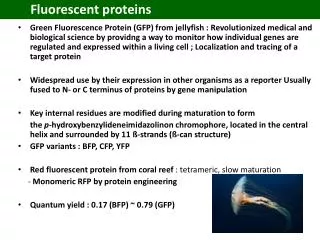
Fluorescent proteins. Green Fluorescence Protein (GFP) from jellyfish : R evolutionized medical and biological science by providing a way to monitor how individual genes are regulated and expressed within a living cell ; Localization and tracing of a target protein

E N D
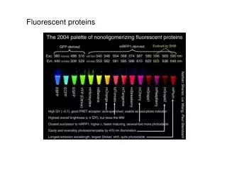
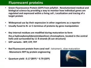
Fluorescent proteins • Green Fluorescence Protein (GFP) from jellyfish : Revolutionized medical and biological science by providing a way to monitor how individual genes are regulated and expressed within a living cell ; Localization and tracing of a target protein • Widespread use by their expression in other organisms as a reporter • Usually fused to N- or C-terminus of proteins by gene manipulation • Key internal residues are modified during maturation to form the p-hydroxybenzylideneimidazolinonchromophore, located in the central helix and surrounded by 11 ß-strands (ß-can structure) • GFP variants : BFP, CFP, YFP • Red fluorescent protein from coral reef : tetrameric, slow maturation - Monomeric RFP by protein engineering • Quantum yield : 0.17 (BFP) ~ 0.79 (GFP)
History of Fluorescent Proteins • 1960s : Curiosity about what made the jellyfish Aequoreavictoria glow Green protein was purified from jellyfish by Osamu Shimomura in Japan. • Utility as a tool for molecular biologists was not realized until 1992 when Douglas Prasher reported the cloning and nucleotide sequence of wt-GFP in Gene. - The funding for this project had run out, and Prasher sent cDNA samples to several labs. • 1994 : Expression of the coding sequence of fluorescent GFP in heterologous cells of E. Coli and C. elegansby the lab of Martin Chalfie : published in Science. • Although this wt-GFP was fluorescent, it had several drawbacks: dual peaked excitation spectra, poor photo-stability, and poor folding at 37°C.
1996 : Crystal structure of a GFP Providing a vital background on chromophore formation and neighboring residue interactions. Researchers have modified these residues using protein engineering (site directed and random mutagenesis) Generation of a wide variety of GFP derivatives emitting different colors ; CFP, YFP, CFP by Roger Y. Tsiengroup Applications in many areas including cell biology, drug discovery, diagnostics, genetics, etc. • 2008 : Martin Chalfie, Osamu Shimomura and Roger Y. Tsien shared the Nobel Prize in Chemistry for their discovery and development of the fluorescent proteins.
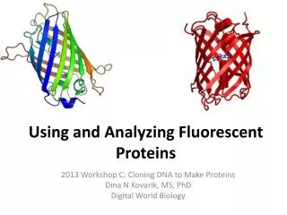
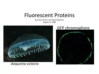
GFP (Green Fluorescent Protein) • Jellyfish Aequoreavictoria • A tightly packed -can (11 -sheets) enclosing an -helix containing the chromophore • 238 amino acids • Chromophore • Cyclic tripeptide derived from Ser(65)-Tyr(66)-Gly(67) • Wt-GFP absorbs UV and blue light (395nm and 470nm) and emits green light (maximally at 509nm)
GFP and chromophore • Covalently bonded chromophore : 4-(p-hydroxybenzylidene)imidazolidin-5-one (HBI). • HBI is nonfluorescent in the absence of the properly folded GFP scaffold and exists mainly in the • unionized phenol form in wt-GFP. • Maturation (post-translational modification) : Inward-facing side chains of the barrel induce • specific cyclization reactions in the tripeptide Ser65–Tyr66–Gly67 that induce ionization of HBI to • the phenolate form and chromophore formation. • The hydrogen-bonding network and electron-stacking interactions with these side chains influence • the color, intensity and photo-stability of GFP and its numerous derivatives
Diverse Fluorescent Proteins by Protein Engineering wtGFP : Ser(65)-Tyr(66)-Gly(67)
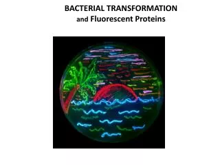
Fluorescence emission by diverse fluorescent Proteins The diversity of genetic mutations is illustrated by this San Diego beach scene drawn with living bacteria expressing 8 different colors of fluorescent proteins.
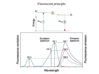
Absorption and emission spectra a) Normalized absorption and b) fluorescence profiles of representative fluorescent proteins: cyan fluorescent protein (cyan), GFP, Zs Green, yellow fluorescent protein (YFP), and three variants of red fluorescent protein (DS Red2, AS Red2, HC Red). From Clontech.