Download
1 / 11
110 likes | 237 Views
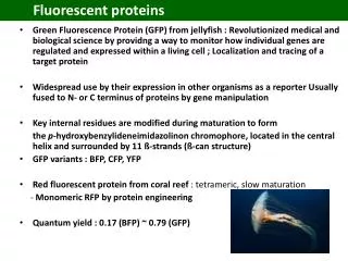
Fluorescent Proteins By James Dicarlo and Ingrid Spielman August 12, 2008. GFP chromophore. Aequorea victoria. Tons of different Fluorescent Proteins . Shape. Discosoma is a coral. Conjugated Double Bonds. p-hydroxybenzylidene-imidazolidone. Stokes Shift. Conjugated double bonds:

E N D
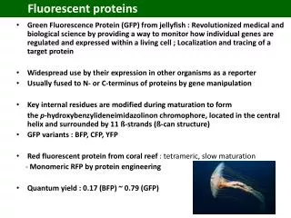
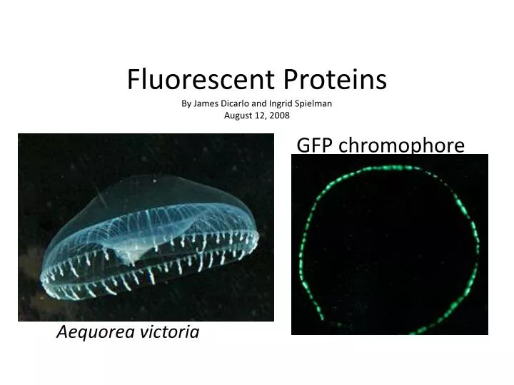
Fluorescent ProteinsBy James Dicarlo and Ingrid SpielmanAugust 12, 2008 GFP chromophore Aequorea victoria
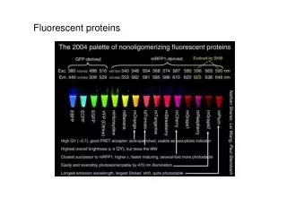
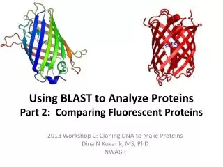
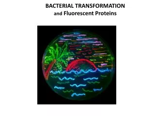
Tons of different Fluorescent Proteins Shape Discosoma is a coral
Conjugated Double Bonds p-hydroxybenzylidene-imidazolidone Stokes Shift • Conjugated double bonds: • Ring Structure • Pi bonds • The more the merrier: lower energy photons required (visible light) • Quantum efficiency increases as number of pi bonds increase • To fall back to Ground Level State: • Vibrational relaxation • Rotational energy • Emission
IDEAL FPs? • Sufficient signaling: Brightness measured by quantum yield; the ratio of photons absorbed to photons emitted • High photostability: does not bleach out easily • Minimal cross-talk between excitation and emission • Fold in vivo at certain pH and temperature levels • Should not oligomerize: can engineer into monomers via A206K mutation • Not toxic
Our FPs • GFP 395 /509 nm • mCherry 587/610 • Venus YFP 502/532 • Sapphire 399/508
A Rabbit or Goat antibody is created to • the antigen of interest • 2) A secondary antibody is based on • the primary antibody. • 3) The Primary antibody is bound • to the antigen of interest and then • the secondary (dye coupled) antibody • is used to locate the primary antibody. • Pros-This method is useful for locating different cellular bodies but use the same secondary dye coupled antibody. • Cons- This method interferes with cell function while fusion GFP constructs allow for viewing of intact cell/macromolecule • http://www.bio.davidson.edu/COURSES/genomics/method/IMF.html Immunofluorescence
FRET • Fluorescence Resonance Energy Transfer • Emission wavelength of one fluorescent protein (or other chromophore) serves as Absorption wavelength for other Protein http://en.wikipedia.org/wiki/Fluorescence_resonance_energy_transfer
FACS • Fluorescence Activated Cell Sorting • Laser is passed through flowing liquid to excite chromophores of a Specific wavelength. • Can determine density and composition of mixture • Multiple lasers can be used to separated different populations of cells http://www.invitrogen.com/site/us/en/home/support/Tutorials.html