Download
1 / 55
550 likes | 737 Views
Partnerships for Success: Corneal Transplant in the 21st Century. Presenters: Christopher Blanton, MD Medical Director, Ocular Services, OneLegacy Jessica Horton Respiratory Therapist & Cornea Recipient, Antelope Valley Hospital Moderator:

E N D
Partnerships for Success: Corneal Transplant in the 21st Century Presenters: Christopher Blanton, MDMedical Director, Ocular Services, OneLegacy Jessica HortonRespiratory Therapist & Cornea Recipient, Antelope Valley Hospital Moderator: Sherri Lamon, RN, PIH Health Breakout Session A
Objectives • Understand the importance of good eye care • Recognize the benefits of timely referrals • Be aware of indications for corneal transplant and innovative remedies.
Question to Run on • How do your actions in the hospital affect the gift of sight?
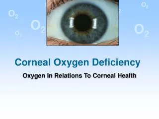
The Basics The goal of the cornea is to focus light to a point on the retina.
What two requirements are necessary for the cornea to perform this function? Transparency and correct shape
Corneal Anatomy • Epithelium- 50 microns • Bowman’s Layer- 12 microns • Stroma- 450-900 microns • Descemet’s membrane- 3-12 microns • Endothelium- 4-6 microns
Pseudophakic Bullous Keratopathy
Corneal Dystrophy Treated with Excimer Laser
Partial Thickness • DLEK • DSEK • DSAEK
Deep Lamellar • Replacing Descemet’s and Endothelial layer • Difficult dissection technique for both donor and host cornea
Descemet’s Stripping • Manual Dissection-host and donor • Automated -donor
IntraLase Enabled Keratoplasty The IntraLase Enabled Keratoplasty software can be programmed to produce different configurations with computer precision. Historical use of trephination utilizing different shapes has taught us that different configurations have unique features that can be used for specific indications.
IntraLase Enabled Keratoplasty The IntraLase® FS ophthalmic surgical laser is cleared for the creation of a lamellar cut/resection of the cornea for lamellar keratoplasty and for the creation of a PENETRATING CUT/INCISION for penetrating keratoplasty The IntraLase FS surgical laser can perform the following cuts that when combined at different angles can create various geometrically shaped corneal resections at the surgeon’s desired depth.
IntraLase Enabled Keratoplasty The IntraLase Enabled Keratoplasty application allows the user to perform three cut segments: a posterior side cut, lamellar cut, and anterior side cut.
The IntraLase Enabled Keratoplasty application allows the user to perform three cut segments: a posterior side cut, lamellar cut, and anterior side cut.
Cut Angles Red 90 degrees Blue 30 degrees Yellow 150 degrees Dotted line angle measurement
Key Elements in Caring for Potential Donors • Making the Referral • Approach for Donation • Maintain the Opportunity • Ventilated or Comatose Patients
Making the Referral • Report all imminent/cardiac deaths within ONE HOUR • Regardless of medical condition, age, advanced directive • HIPAA compliant
Why? • Allows time to serve the family • Preserves organ and tissue viability • Ensures compliance with federal and state laws
Approaching for Donation • Most deaths will not be eligible for donation • Do not approach independently of OneLegacy. • Have medical chart available for OneLegacy to determine suitability. • LNOK contact information
Maintain Opportunity for Corneal Donation • Ventilated or comatose patients- lubricate eyes frequently : every 4-6 hours gel is better than tears • Deceased patients- wait for go ahead from One Legacy: eye care may interfere with M.E./coroner's investigation.