Download
1 / 18
190 likes | 813 Views
Neurons and Glia. Chapter 2 Pg 32-57. Obstacles to Study. Cells are too small to see. To study brain tissue with a microscope, thin slices are needed but the brain is like jello. Formaldehyde used to “fix” or harden tissue early in 19 th century. Brain tissue is all the same color:

E N D
Neurons and Glia Chapter 2 Pg 32-57
Obstacles to Study • Cells are too small to see. • To study brain tissue with a microscope, thin slices are needed but the brain is like jello. • Formaldehyde used to “fix” or harden tissue early in 19th century. • Brain tissue is all the same color: • Nissl stain revealed cell bodies – cytoarchitecture • Golgi stain revealed parts of the neuron.
Brodmann Areas • Different areas of the brain with different functions have different kinds of neurons. • Brodmann mapped the areas based on the kinds of cells found: • Cytoarchitectonic method • 52 functionally distinct areas identified by number.
Ramon y Cajal’s Principles • Neuron doctrine – neurons are like other cells. • Principle of dynamic polarization – electrical signals flow in only one, predictable direction within the neuron. • Principle of connectional specificity: • Neurons are not connected to each other, but are separated by a small gap (synaptic cleft). • Neurons communicate with specific other neurons in organized networks – not randomly.
Neuronal Circuits • Neurons send and receive messages. • Neurons are linked in pathways called “circuits” • The brain consists of a few basic patterns of circuits with many minor variations. • Circuits can connect a few to 10,000+ neurons.
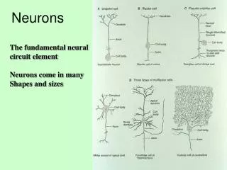
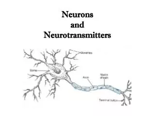
Parts of the Neuron • Soma – the cell body • Neurites – two kinds of extensions (processes) from the cell: • Axon • Dendrites • All parts of the cell are made up of protein molecules of different kinds.
How Neurons Communicate • An all-or-nothing electrical signal, called an action potential, travels down the axon. • The amplitude (size) of the action potential stays constant because the signal is regenerated. • The speed of the action potential is determined by the size of the axon. • Action potentials are highly stereotyped (very similar) throughout the brain. • At the end of the axon (terminal button), neurotransmitter is released, which may start an action potential in another neuron.
The Synapse • The synapse is the point of contact between neurons. • Axon terminal button makes contact with some part of an adjacent neuron. • Synaptic vesicles containing neurotransmitter open when there is an action potential. • Neurotransmitter may enter the adjacent neuron – unused neurotransmitter is reabsorbed (reuptake).
Dendrites • Dendrites function as the antennae of the neuron, receiving input from other neurons. • Dendrites are covered with synapses. • Each synapse has many receptors for neurotransmitters of various kinds. • Dendritic spines – specialized dendrites that isolate reactions at some synapses.
How to Tell Axons from Dendrites • Dendrites receive signals – axons send them. • There are hundreds of dendrites but usually just one axon. • Axons can be very long (> 1 m) while dendrites are < 2 mm. • Axons have the same diameter the entire length – dendrites taper. • Axons have terminals (synapses) and no ribosomes. Dendrites have spines (punching bags). • Don’t be fooled by the branches – both have them.
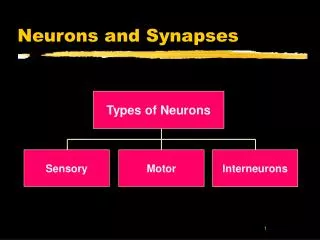
Ways of Classifying Neurons • By the number of neurites (processes): • Unipolar, bipolar, multipolar • By the type of dendrites: • Pyramidal & stellate (star-shaped) • By their connections (function) • Sensory, motor, relay interneurons, local interneurons (Golgi Type II neurons) • By neurotransmitter – by their chemistry
Parts of the Soma (Cell Body) • Nucleus – stores genes of the cell (DNA) • Organelles – synthesize the proteins of the cell • Cytosol – fluid inside cell • Plasmic membrane – wall of the cell separating it from the fluid outside the cell.
Organelles • Mitochondria – provide energy • Microtubules – give the cell structure • Rough endoplasmic reticulum – produces proteins needed to carry out cell functioning • Ribosomes – produce neurotransmitter proteins • Smooth endoplasmic reticulum – packages neurotransmitter in synaptic vesicles • Golgi apparatus – Part of the smooth endoplasmic reticulum that sorts proteins for delivery to the axon and dendrites
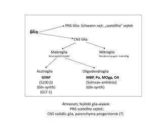
Kinds of Cells • Neurons (nerve cells) – signaling units • Glia (glial cells) – supporting elements. • Miscellaneous other cells: • Ependymal cells – form the lining of the ventricles, also aid brain development • Microglia – remove debris left by dead or degenerating neurons and glia. • Veins, arteries, and capillaries in the brain.
Functions of Glia • Separate and insulate groups of neurons • Produce myelin for the axons of neurons • Scavengers, removing debris after injury • Buffer and maintain potassium ion concentrations • Guide migration of neurons during development • Create blood-brain barrier, nourish neurons
Kinds of Glia • Oligodendrocytes – surround brain & spinal cord neurons and give them support. • In white matter, provides myelination • In gray matter, surround cell bodies • Schwann cells – provide the myelin sheath for peripheral neurons (1 mm long). • Astrocytes – absorb potassium, perhaps nutritive because endfeet contact capillaries (blood vessels), form blood-brain barrier.