Neurons
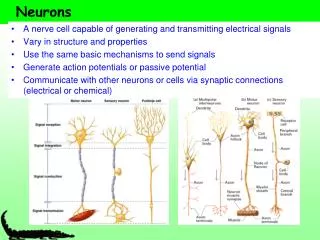
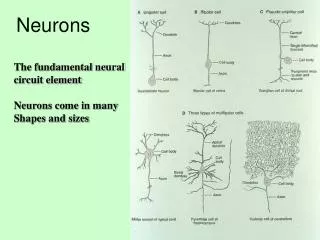
Neurons. The two principal cell types of the nervous system are: Neurons – excitable cells that transmit electrical signals. There are 150 different types. Vary in function and size. Supporting cells (neural glia)– cells that surround and wrap neurons.

Neurons
E N D
Presentation Transcript
Neurons • The two principal cell types of the nervous system are: • Neurons – excitable cells that transmit electrical signals. • There are 150 different types. • Vary in function and size. • Supporting cells (neural glia)– cells that surround and wrap neurons. • Oligodendrocytes, Schwann cell, Astrocytes
Supporting Cells: Neuroglia • The supporting cells • Provide a supportive scaffolding for neurons • Segregate and insulate neurons • Guide young neurons to the proper connections • Promote health and growth
Fundamental Types of Neurons • Sensory (afferent) neurons • detect changes in body and external environment • information transmitted to brain or spinal cord • Interneurons (association neurons) • lie between sensory and motor pathways in CNS • 90% of our neurons are interneurons • Integration and retrieving information • Motor (efferent) neuron • send signals out to muscles and gland cells • organs that carry out responses called effectors
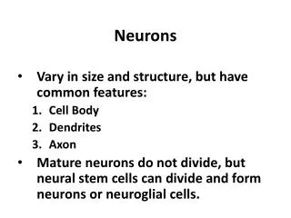
Structure of a Neuron • Cell body (soma) • single, central nucleus • contains many multi-branched dendrites • Which receive signals from other neurons. • Axon • (nerve fiber) arising from axon hillock for rapid conduction • Axon terminals release will neurotransmitters that communicate a chemical message to another nerve or muscle
Neuron Physiology • Living nerve cells are polarized • The inside of the cell (Intracellular fluid) ICF is negatively charged compared to the outside of the cell extracellular fluid (ECF) • The cell is able to maintain a resting membrane potential of -70 mV (negative charge on the inside of membrane by active transport and specific voltage gated channels.
Resting Membrane Potential The cell membrane is considered a semi-permeable membrane which selectively allows things in and out of the cell. • Large negatively charged molecules found in the ICF such as proteins and phosphates are confined to the inside of the cell. The membrane is impermeable to these molecules which contributes to the ICF maintaining RMP of -70mV
Ionic Basis of Resting Membrane Potential • Na+ concentrated outside of cell (ECF) • K+ concentrated inside cell (ICF)
Basis of the Resting Membrane Potential • Since Na+ ion are more concentrated in the ECF when a specific voltage gated Na+ channel opens Na+ will always rush into the cell by diffusion. • Since K+ ion channels are more concentrated in the ICF when a specific voltage gated K+ channel opens K+ will always rush out of the cell by diffusion • In order to keep the resting membrane potential at –70 mV the cell is constantly hydrolyzing ATP with the Na+,K+-ATPase pump.
Action Potentials (APs) • There are 3 phases to an AP: • Depolarization • a reduction in the polarity of the membrane potential by allowing Na+ to enter the cell. • Repolarization • membrane potential returns towards the resting value closing Na channels and opening K+ channels. K+ travels along its concentration gradient out of the cell returning the inside of the cell to a negative value. • Hyperpolarization • Slow closing K+ channels cause the inside of the cell to be more negative than the resting value • All APs have the same magnitude regardless of the size of the stimulus
The Refractory Period • Absolute refractory period • as long as Na+ gates are open • no stimulus will trigger AP • Relative refractory period • as long as K+ gates are open • only especially strong stimulus will trigger new AP • Refractory period is occurring only to a small patch of membrane at one time (quickly recovers)
Impulse Conduction in Unmyelinated Fibers • The action potential in trigger zone begins impulse • Nerve signal (impulse) - a chain reaction of sequential opening of voltage-gated Na+ channels down entire length of axon • This is a very slow process. (2 m/sec) • We need something to speed the process up.
Saltatory Conduction - Myelinated Fibers • The velocity of an action potential propagates along the length of the axon depends on: • axon diameter • The larger the diameter of the axon the greater the velocity of the action potential travels along the axon to the axon terminal. • Myelin sheath increases the diameter of sections of the axon which dramatically increases impulse speed. (120 m/sec)
Myelin Sheath • Myelin is a white, fatty insulating covering around most of the long axons. It plays an important role in both conduction velocity and protection of the axon. • In the CNS Oligodendrocytes can myelinate many different neurons. • In the PNS Schwann cells are can only myelinate a portion of one axon.
Diseases of the Nervous System • What would be worse? • A disease that attacks neurons of the CNS • A disease that attacks neurons of the PNS • What are the deficits one might expect to see if the neurons loose their Myelination?
Saltatory Conduction • Notice how the action potentials jump from node of Ranvier to node of Ranvier.
Presynaptic Neurons • Presynaptic neurons • Nerve signal(AP) opens voltage-gated calcium channels allowing it to diffuse into the synaptic knob. • Calcium triggers the release of a neurotransmitter such as acetylcholine (Ach) from the (synaptic vesicles). • The neurotransmitters are released into the synaptic cleft.
Postsynaptic Neuron • Neurotransmitters diffuse across the synaptic cleft binding to ligand-gated channels on the postsynaptic neuron. • Graded Potential • Specific neurotransmitters can influence the permeability of the voltage gated channels. • This influences the post synaptic neuron to become more likely to generate an AP (depolarization) or less likely (hyperpolarization).
Postsynaptic Potentials- EPSP • Excitatory postsynaptic potentials (EPSP) • a positive voltage change causing postsynaptic cell to be more likely to fire • result from Na+ flowing into the cell • glutamate and aspartate are excitatory neurotransmitters
Postsynaptic Potentials- IPSP • Inhibitory postsynaptic potentials (IPSP) • a negative voltage change causing postsynaptic cell to be less likely to fire (hyperpolarize) • result of Cl- flowing into the cell or K+ leaving the cell • glycine and GABA are inhibitory neurotransmitters
The membrane potential of a real neuron typically undergoes many EPSPs (A) and IPSPs (B), since it constantly receives excitatory and inhibitory input from the axons terminals that reach it.
Summation - Postsynaptic Potentials • Net postsynaptic potentials in trigger zone • firing depends on net input of other cells • The trigger zone takes in both EPSP and IPSPs. If there are more EPSPs threshold will be reached. • temporal summation • single synapse receives many EPSPs in short time • spatial summation • single synapse receives many EPSPs from many cells
Summation of EPSP’s • Does this represent spatial or temporal summation?
Action Potentials vs. Graded Potentials • Action Potentials (All-or-none phenomenon) • action potentials either completely, or not at all • Graded Potentials ( sub threshold) • EPSP and IPSPs are • are graded (vary in magnitude with stimulus strength) • are decremental (get weaker the farther they spread) • are reversible as K+ diffuses out of cell
Clinical Applications • Many drugs work by altering neuronal functions. • Block the receptor site • Beta blockers: prevent sympathetic input to the heart and various organs • Block the reabsorption of the neurotransmitter • SSRI: Prevent the reuptake of serotonin so it stays in the synaptic cleft longer and continue to stimulate nerve • Bind to the receptor site • Curare blocks the (ACh) acetylcholine receptors by binding to the same position on the receptor