Download
1 / 21
260 likes | 1.1k Views
NEURONS AND GLIA. Introduction. “Neurophilosophy” Brain (neurons) is the origin of mental abilities Glia and Neurons Glia Insulates, supports, and nourishes neurons 90% of brain cells are glial cells Neurons Process information Sense environmental changes

E N D
Introduction • “Neurophilosophy” • Brain (neurons) is the origin of mental abilities • Glia and Neurons • Glia • Insulates, supports, and nourishes neurons • 90% of brain cells are glial cells • Neurons • Process information • Sense environmental changes • Communicate changes to other neurons • Command body response
The Neuron Doctrine • The birth of neurohistology • Microscopy invention • Discovery of fixation method for cutting thin slices • Staining methods for selectively coloring parts of cells • The Nissl Stain • Developed by by the German neurologist Franz Nissl • Stain the nuclei and surrounding material (Nissl body) • Made it possible to distinguish neurons vs. glia and to study the arrangements of neurons in different parts of brain (cytoarchitecture) Fig2.1
The Neuron Doctrine • The Golgi Stain • Camillo Golgi discovered that by soaking brain tissue in a silver chromate solution, a small percentage of neurons became darkly colored in their entirety • Soma (cell body or perikaryon) and neurites (axons and dendrites) Fig 2.3
The Neuron Doctrine • Cajal’s Contribution • Santiago Ramon y Cajal used Golgi stain method and worked out the circuitry of many regions of the brain : Father of neuroanatomy • Golgi versus Cajal • Reticular theory vs. cell theory • Neuron doctrine • Neurons communicate by contact, not continuity • Final proof had to wait until EM got developed in the 1950s
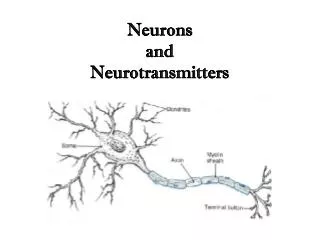
The Prototypical Neuron • The Soma • ~20 um in diameter • Cytosol: Potassium rich watery fluid inside the cell • Organelles: Membrane-enclosed structures within the soma • Cytoplasm: Contents within a cell membrane (e.g., organelles, excluding the nucleus) Fig2.7
The Prototypical Neuron • The Axon • Begins with axon hillock, initial tapered segment where action potentials are generated • Rough ER does not extend into axon • Protein composition of axon membarane is fundamentally different from that of soma • No protein synthesis in the axon • May extend from less than a millimeter to over a meter long • May branch out (generally at right angles) to form axon collaterals that could return to the same cell (recurrent collaterals) • Diameter ranges from less than 1 mm to 25 mm in humans - The speed of nerve impulses depends on axonal diameter
The Prototypical Neuron The Axon Terminal (terminal bouton) • A site where the axon comes in contact with other neurons and passes information on to them • Terminal arbor or boutons en passant • Synapse - To fasten together • Innervation - making synaptic contact • Differences between the cytoplasm of axon terminal and that of axon • No microtubules in the terminal • Presence of synaptic vesicles (~50 nm in diameter) • Dense covering of proteins on the inside surface of the synaptic membrane • Large number of mitochondria (high energy demand)
The Prototypical Neuron Synapse • Pre- and Postsynaptic sides : directionality of information flow • Synaptic transmission • Synaptic cleft • Electrical-to-chemical-to-electrical transformation • Neurotransmitter
The Prototypical Neuron • Axoplasmic transport • Wallerian degeneration • Degeneration of axon when severed (axotomy) is due to the lack of protein synthesis machinery within axon Kandel Fig 55-18 • Anterograde transport by kinesin and retrograde transport by MAP-1C (dynein)
The Prototypical Neuron • Slow Axoplasmic transport • Paul Weiss’s experiment • Tied off a sciatic nerve (axon) to find that material accumulate on the proximal side of the knot • When the knot was untied, the bulged out accumulation continued down the axon • The speed of movement was measured to be about 1 - 10 mm per day ; SLOW AXOPLASMIC TRANSPORT • Only anterograde direction • Slow transport itself can be at two different speeds • Slower (0.2-2.5mm per day) : fibrillar elements of cytoskeleton (neurofilament subunits, tubulins..) • Faster (about twice as fast as the slower) : various cytosolic proteins (clathrin, actin, actin-binding proteins, enzymes..)
The Prototypical Neuron • Fast Axoplasmic transport • Bernice Grafstein • Injected radioactive amino acids into somata • Traced the synthesized (hot) proteins along the axon • Large membraneous organelles are transported via fast transport • Includes vesicles of the constitutive secretory pathways, synaptic vesicles precursor membranes, mitochondria, smooth ER elements.. • ATP dependent but not protein synthesis dependent (once synthesized) • Soma-independent (isolated axon still can transport
The Prototypical Neuron • Dendrites • Greek for ‘tree’ • Dendritic tree for all the dendrites of a neuron • “Antennae” of neurons - covered with thousands of synapses • Dendritic membrane (postsynaptic membrane) contains many specialized receptors for neurotransmitters • Dendritic spines • Some neurons have these structures for receiving some types of inputs • Discovered by Cajal • Believed to isolate various chemical reactions • Dynamic structures affected by the type and amount of inputs and developmental changes of environment Fig 2.17 Fig 2.18
Mental Retardation and dendritic spines • Brain function depends on the highly precise synaptic connections, which are formed during the fetal period and are refined during infancy and early childhood • 95% of population falls within two standard deviations from the mean of IQ (around 70 when the mean is set to be 100). Some 2-3% of humans with intelligence score below are considered to be mentally retarded IF the cognitive impairment affects the person’s ability to adapt their behavior to the setting in which they live • Can have many causes • Genetic disorders such as PKU or Downsyndrome • Accidents or infection during pregnancy or early childhood • Poor nutrition during pregnancy • Environmental impoverishment such as the lack of good nutrition, socialization, sensory stimulation during infancy • Some with clear physical correlates (retarded growth, abnormal structures of head, hands, and body), most with only behavioral manifestations • Dendritic spine abnormality has been found to be correlated with mental retardation Fig A
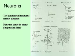
Classifying Neurons • Classification Based on the Number of Neurites • Unipolar cell • Found in invertebrate nervous system - single process with different segments serving as receptive surfaces or releasing terminals • Bipolar cell • Two neurites • Multipolar cell • Most neurons in the brain are multipolar Kendal fig 2-4
Classifying Neurons • Classification Based on Dendritic and Somatic Morphologies • Often unique to a particular region of the brain • Cortex - Stellate cells (star-shaped) and pyramidal cells (pyramid-shaped) • Spiny or aspinous
Classifying Neurons • Further Classification • Based on connections within the CNS • Primary sensory neurons • motor neurons • interneurons • Based on axonal length • Golgi Type I - projection neurons that extend their axons to other parts of the brain (e.g. pyramidal neurons in the cortex) • Golgi Type II - local circuit neurons that have short axons that do not extend beyond the vicinity of cell body (e.g. stellate cells in the cortex) • Based on neurotransmitter type • Cholinergic, glutamatergic, GABAergic…
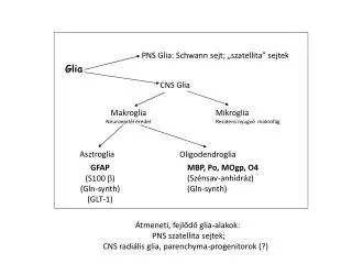
Glia • ‘Sleeping Giants’ ? • Function of Glia • Supports neuronal functions • Without glia brain cannot function! • Astrocytes • Most numerous glia in the brain • Fill spaces between neurons • Imporatant regulator of the chemical contents of extracellular spaces (Not much left after filling up) • Envelop synaptic junctions - restrict the spreading of released neurotransmitters • Possess their own neurotransmitter receptors!!
Glia • Myelinating Glia • Oligodendroglia (in CNS) and Schwann cells (in PNS) • Insulate axons by wrapping axons around • Myelin sheath • One Oligodedroglia can provide insulation to several axons but each Schwann cell does to a only a single axon • Node of Ranvier • Region where the axonal membrane is exposed
Glia • Other Non-Neuronal Cells • Microglia as phagocytes (immune) • Ependymal cells provide lining of fluid-filled ventricles and directs cell migration during brain development • Vasculature