Download
1 / 12
120 likes | 434 Views
Proprioception and Discriminative Touch – Dorsal Column/Medial Lemniscus System. I. Sensory Systems.

E N D
Proprioception and Discriminative Touch – Dorsal Column/Medial Lemniscus System
I. Sensory Systems • Physicochemical changes (stimuli) in the outer or inner environments of the individual are detected by morphologically diverse types of receptors. Sensation is the ability of the body to detect a stimulus. • Modality (type of sensation, eg. Pain, temperature, touch, sound, etc) is determined by stimulus specificity and properties of receptors. • Modalities are represented along specific neuron chains (aka nerve cell pathways). • Frequency of impulses along nerve fibers determine intensity of sensation. • Location of stimulus is maintained along nerve cell pathways (topographic representation).
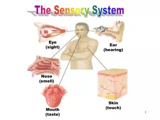
II. Somatic Sensations • Discriminative touch. Usually tested as two-point discrimination. Detected by Meisner’s corpuscles, Merkel discs, peritrichial endings and free nerve endings. • Pressure and vibration. Tested by tuning fork. Detected by Pacinian and Meisner’s corpuscles. • Light (crude) touch. Tested with wisp of cotton. Detected by peritrichial and free nerve endings. • Pain and temperature. Tested by pinprick of thermal application. Detected by free nerve endings and some encapsulated receptors. • Proprioception. Conscious. Tested by joint movement. Detected by diffuse endings in joints and joint capsules. Unconscious. Tested by muscle tone and gait assessment. Detected by muscle spindles and Golgi tendon organs
VI. Column – Medial Lemniscus Dorsal System • Mediates proprioceptive (both conscious and unconscious), discriminative touch, deep pressure and vibratory senses. • Cell body of 1st order neuron is in the dorsal root ganglion (DRG). • Peripheral process of DRG cell terminates as or is incorporated into receptor. • Central process enters into dorsal column of spinal cord by way of med div of dorsal root and ascends in the ipsilateral fasciculus gracilis or fasciculus cuneatus. • F. gracilis and f. cuneatus terminate onto a 2nd order neuron in the nuclei gracilis and cuneatus at the level of the caudal medulla. • Collaterals of the ascending fibers are used for reflex activity.
VI (cont.) Somatotopic organization in the F. gracilis and F. cuneatus • F. gracilis contains fibers representing ipsilateral dermatomes S5-T6. F. cuneatus represents dermatomes T6-C2. • F. gracilis contains representation of lower limb, abdomen and lower thorax. F. cuneatus represents upper thorax, upper limb and back of head. • F. cuneatus appears at T6 spinal segment and above that level is separated from F. gracilis by the posterior intermediate septum and sulcus. • Unilateral lesions in F. gracilis or cuneatus results in loss of proprioception, two-point discrimination, vibratory sense and deep pressure in all ipsilateral dermatomes below the lesion level.
VI. (Cont.) 2nd Order Neuron • Cell body localized in the nucleus gracilis or nucleus cuneatus at lower medulla level. • Axon leaves n. gracilis or cuneatus, course ventrally and medially in the internal arcuate fibers,crosses the midline and ascends in the medial lemniscus (ML) just dorsal to the medullary pyramid (pyramidal tract). • Fibers representing sacral dermatomes are located most ventrally in the ML. Fibers representing cervical dermatomes are located most dorsally in the ML. • Long axis of ML rotates in the pons with the ML assuming a horizontal orientation. At this level the ALS joins the ML laterally. • Throughout the rostral midbrain the ML is dorsal and lateral to the red nucleus. • Axons of the ML terminate in ventral posterolateral (VPL) nucleus of the thalamus. • Unilateral lesions of ML throughout its course in the brainstem causes the loss of proprioception, discriminatory touch and vibratory sense over contralateral dermatomes C2-S5.
VI. (Cont.) 3rd Order (thalamocortical) Neuron • Cell body of 3rd order neuron is located in the VPL nucleus of the thalamus. • Axon of 3rd order neurons leaves VPL, enters ipsilateral internal capsule and ascends to the somatosensory (Brodmann’s areas 3,1,2) cortex in the postcentral gyrus. • Termination of fibers is such that a “sensory homonculus” representing the contralateral dermatomes is formed over the somatosensory cortex. • Different modalities are represented in different portions of 3,1,2. • 3,1,2 projects to a secondary somatosensory (SII) cortex and to 5,7. Processing in 5 and 7 results in appreciation of finer qualities of the modality (e.g. recognition of shape by tactile discrimination or stereognosis).
VI. Clinical Correlation • Romberg’s sign. Lesions in dorsal column – ML system causes the loss of conscious proprioception (position sense). Ask patient to stand upright with eyes closed. If patient is positive for Romberg, he/she sways from side to side and may fall. • Tabes dorsalis – Destruction of dorsal columns due to tertiary syphyllis (neurosyphillis). Often f. gracilis is involved bilaterally. Patient has wide-based, staggering gait, often accompanied by slapping feet against the ground when walking. This lesion will be discussed again in the lecture on spinal cord lesions. • Unilateral lesions in dorsal column produce ipsilateral signs. Unilateral lesions in the ML produce signs contralaterally. Unilateral VPL or 3,1,2 lesions produce contralateral signs.