Download
1 / 40
400 likes | 516 Views
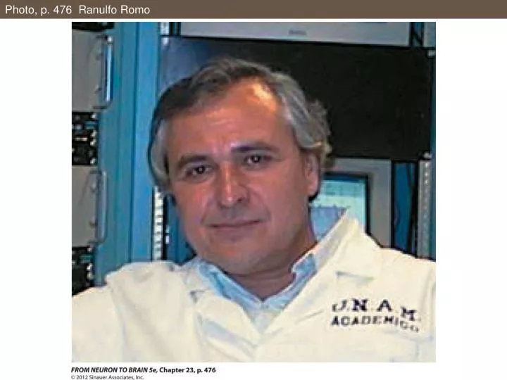
Photo, p. 476 Ranulfo Romo. Figure 23.1 Vibration Discrimination Task and Performance. Figure 23.1 Vibration Discrimination Task and Performance (Part 1). Figure 23.1 Vibration Discrimination Task and Performance (Part 2).

E N D
Figure 23.1 Vibration Discrimination Task and Performance (Part 1)
Figure 23.1 Vibration Discrimination Task and Performance (Part 2)
Figure 23.2 Neuronal Coding of Vibration Frequency in Primary Somatosensory Cortex (SI)
Figure 23.2 Neuronal Coding of Vibration Frequency in Primary Somatosensory Cortex (SI)(Part 1)
Figure 23.2 Neuronal Coding of Vibration Frequency in Primary Somatosensory Cortex (SI)(Part 2)
Figure 23.3 Replacement of Vibrations by Artificial Stimuli (Part 1)
Figure 23.3 Replacement of Vibrations by Artificial Stimuli (Part 2)
Figure 23.3 Replacement of Vibrations by Artificial Stimuli (Part 3)
Figure 23.4 Neuronal Activity in SI Cortex Characterized by Firing-Rate Coefficients a1 and a2
Figure 23.5 Cortical Regions Involved in the Vibration Comparison Task
Figure 23.6 Neuronal Activity beyond SI Characterized by Firing Rate Coefficients a1 and a2
Figure 23.6 Neuronal Activity beyond SI Characterized by Firing Rate Coefficients a1 and a2(Part 1)
Figure 23.6 Neuronal Activity beyond SI Characterized by Firing Rate Coefficients a1 and a2(Part 2)
Figure 23.7 Time Course of Response Parameters throughout the Trial Period
Figure 23.7 Time Course of Response Parameters throughout the Trial Period (Part 1)
Figure 23.7 Time Course of Response Parameters throughout the Trial Period (Part 2)
BOX 23.1 Functional Magnetic Resonance ImagingBrain Activity as Revealed by Averaging fMRI Scans across a Group of 20 Subjects
Figure 23.10 Brain Activation Directly Correlated with Face Perception
Figure 23.11 Responses of Macaque Monkey Inferotemporal Neurons to Changes in Viewing Angle and Stimulus Size
Figure 23.11 Responses of Macaque Monkey Inferotemporal Neurons to Changes in Viewing Angle and Stimulus Size (Part 1)
Figure 23.11 Responses of Macaque Monkey Inferotemporal Neurons to Changes in Viewing Angle and Stimulus Size (Part 2)
Figure 23.11 Responses of Macaque Monkey Inferotemporal Neurons to Changes in Viewing Angle and Stimulus Size (Part 3)
Figure 23.12 Schematic Organization of M, P, and K Channels to Visual Cortex
Figure 23.12 Schematic Organization of M, P, and K Channels to Visual Cortex (Part 1)
Figure 23.12 Schematic Organization of M, P, and K Channels to Visual Cortex (Part 2)
Figure 23.14 The Direction of Eye Movements Can Be Altered by Electrical Stimulation in Area MT
Figure 23.14 The Direction of Eye Movements Can Be Altered by Electrical Stimulation in Area MT (Part 1)
Figure 23.14 The Direction of Eye Movements Can Be Altered by Electrical Stimulation in Area MT (Part 2)
Figure 23.15 Area MT Microstimulation Affects a Working Memory Task
Figure 23.15 Area MT Microstimulation Affects a Working Memory Task (Part 1)
Figure 23.15 Area MT Microstimulation Affects a Working Memory Task (Part 2)
Figure 23.16 Neuronal Representation of a Concept in Human Hippocampus
Figure 23.16 Neuronal Representation of a Concept in Human Hippocampus (Part 1)
Figure 23.16 Neuronal Representation of a Concept in Human Hippocampus (Part 2)