Download

1 / 21
210 likes | 335 Views
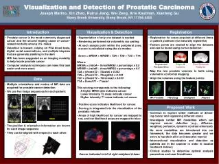
Prostatic Carcinoma. Jakob, 8 yo MC Beagle. History. Diagnosed with prostatic carcinoma 8/26/10 by cytology from ultrasound guided prostatic aspirate done by an internist in Richmond. Originally presented to rDVM for abdominal/hind end pain.

E N D
History • Diagnosed with prostatic carcinoma 8/26/10 by cytology from ultrasound guided prostatic aspirate done by an internist in Richmond. Originally presented to rDVM for abdominal/hind end pain. • Referred to NCSU Oncology for further evaluation & treatment on 8/30/10. Chest radiographs were clear. A focused ultrasound was done for baseline pre-treatment measurements and showed extension of the mass into the urethra. • Chemotherapy protocol of Mitoxantrone and Piroxicam was initiated at the initial visit. • Jakob has received regular recheck chest radiographs and abdominal/focused ultrasounds approximately every 1.5 mo to monitor his disease and response to treatment
Significant Clinical & Laboratory Findings 2/14/11: • Physical Exam • TPR: WNL • CVR: audible low 3rd heart sound (hx of mild MR & TR, mild-mod AR/endocardiosis) • ABD: Mild hepatomegaly • Rectal: Prostate firm and bliaterally enlarged (R>>L), non-painful on palpation, anal sacs normal, normal stool • CBC: WNL
Assesment • Prostate has increased in size <10% = stable disease….. • EXCEPT…… • Thoracic radiographs showed new nodules =progressive disease
Outcome • Jakob is doing well and is not currently exhibiting clinical signs of his disease (no straining to urinate/defecate) • He does progressive disease which means he has “failed” Mitoxantrone chemotherapy. • He was switched to Carboplatin at the 2/14/11 visit
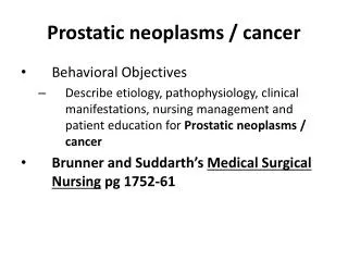
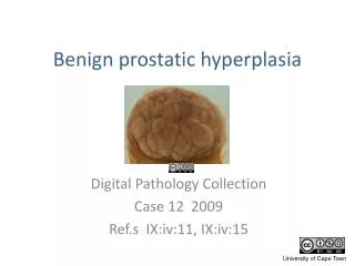
Other Differential Diagnoses • Alternative appearance of Prostatic Carcinoma • Benign Prostatic Hyperplasia • Prostatitis
How do you differentiate? • Signalment • BPH: Intact or recently neutered (within 1 year) • Prostatitis: usually younger males, but can be any age • Neoplasia: usually middle to older animals. High index of suspicion with a large prostate in an older long ago neutered male • Get a sample • Cytology/histopathology needed for a confirmed diagnosis of neoplasia • Prostatic wash with culture to rule in/out prostatitis • Neuter! • If strongly suspect BPH or have ruled out other causes neutering should reduce the size of the prostate in BPH but can take a while to see size reduction