Download

1 / 81
880 likes | 2.38k Views
Allergy and Hypersensitivity. I. Introduction. A. Definitions. Allergy Immune-mediated response to innocuous environmental antigen Can be humoral or cell-mediated reaction Usually involves prior exposure to antigen resulting in sensitization of individual Allergen Innocuous antigen

E N D
A. Definitions • Allergy • Immune-mediated response to innocuous environmental antigen • Can be humoral or cell-mediated reaction • Usually involves prior exposure to antigen resulting in sensitization of individual • Allergen • Innocuous antigen • Universal • Non-reactiving to most people
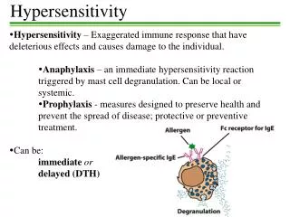
Hypersensitivity reactions • Harmful IRs that cause tissue injury and may cause serious pathologies • Atopy • State of increased susceptibility to immediate hypersensitivity usually mediated by IgE Abs • Over-react to common environmental Ags
B. Four types of immune-mediated hypersensitivity reactions causing tissue damage • Type I = Anaphylaxis hypersensitivity (TH2 = IgE) • Type II = Cytotoxic hypersensitivity (IgG) • Type III = Immune complex hypersensitivity (IgG) • Type IV = Cell-mediated hypersensitivity (TH1, TH2, CTL)
A. Pathway • IgE made during primary response to soluble Ag Binds to high affinity FceRI on mast cells, basophils and activated eosinophils • Sensitizes individual (become allergic) • IgE aka reagin • Secondary exposure allergen binds to IgE on sensitized mast cells, basophils or eosinophils • IgE Ab crosslinking on leads to rapid release of preformed inflammatory mediators
High affinity FceRI is functional on mast cells, basophils, and activated eosinophils. It is composed of a,b and two g chains. Crosslinking of FceRI on cells by Ag and IgE induces degranulation.
Induces degranulation Release of inflammatory mediators [pre-formed substances including histamine, slow reacting substance of anaphylaxis (SRS-A), heparin, prostaglandins, platelet-activating factor (PAF), eosinophil chemotactic factor of anaphylaxis (ECF-A), and various proteolytic enzymes] • Eosinophils release major basic protein which induces degranulation of mast cells and basophils
Tachyphylaxis • Depletion of mast cell granules • Accounts for unresponsiveness of a patient to a skin test following an anaphylactic reaction (lasts 72-96 hours after a reaction)
B. Ig-E mediated reactions differ depending on route of administration and dose • Connective tissue mast cells • Associated with blood vessels • IV-high dose Activated by allergen in the bloodstream systemic • Systemic release of histamine • Systemic anaphylaxis
SC-low dose subcutaneous injection local release of histamine • Wheal and flare reaction
Mucosal mast cells • Inhalation – low dose Activated by inhaled allergen • Smooth muscle contraction of lower airways • Bronchoconstriction • Asthma • Allergic rhinitis (hay fever) • Increased mucosal secretions • Irritations
Fig. 10.24: Allergen-induced release of histamine by mast cells in skin causes localized swelling. Swellings (wheals) appear 20 min. after intradermal injection of ragweed pollen (R), histamine (H). Saline bleb (S) is due to volume of fluid.
Fig. 10.14 Properties of inhaled allergens that favor TH2 priming that promotes IgE isotype switching.
Fig. 10.15 Sensitization to an inhaled allergen. Soluble allergen is processed by APC and displayed to TH2 T cells. T cells help B cells to produce IgE which then binds to mast cells. IL-4 promotes isotype switching to IgE.
Fig. 10.21: Allergic rhinitis (hay fever) is caused by inhaled allergen entering the respiratory tract. Sneezing, runny nose – nasal discharge is full of eosinophils. Allergic conjunctivitis results if the conjunctiva of the eye is affected (itchy, watery, and swelling of eyes).
Ingestion – Activated by ingested allergen • Food allergy • Gut epithelial cells are involved • Intestinal smooth muscle contraction • Vomiting • Diarrhea • Dissemination through bloodstream causes urticaria (hives) or anaphylaxis (rare)
Fig. 10.25: Ingested allergen can cause vomiting, diarrhea and urticaria.
C. Hereditary predisposition for IgE synthesis • FceR genes • Cytokine genes involved in • Isotype switching • Eosinophil survival • Mast cell proliferation • Example: IL-4 promoter mutation which leads to elevated IL-4 can favor IgE • MHC class II • MHC:peptide combinations may favor TH2 response • Example: ragweed pollen associates with HLA-DRB1*1501
D. Type I hypersensitivity reactions can be divided into immediate and late stages • Acute (minutes) versus Chronic (5-12 hours) Reactions • Immediate allergic reactions is then followed by a late-phase response
Acute – Immediate • Peaks within minutes after allergen injection or inhalation and then subsides • Wheal and flare • Bronchial constriction in asthma • IgE crosslinking rapid degranulation • Release of preformed inflammatory mediators • Histamine, serotonin • Mast cell chymase, tryptase, carboxypeptidase and cathepsin G breaks down tissue matrix proteins (remodeling of connective tissue matrix) • TNF-a
Mast cell stained for protease chymase demonstrating abundant granules residing in the cytoplasm.
Chronic – Late • Caused by influx of inflammatory leukocytes (including eosinophils) • Chronic allergic inflammation • Tissue damage • Edema, long-lasting
Chemokines • Heparin • Lipid mediators derived from membrane phospholipids • Form a precursor called arachidonic acid • Many anti-inflammatory agents inhibit arachidonic acid metabolism (e.g. aspirin) • Arachidonic acid forms: • Leukotrienes • Prostaglandins • Thromboxanes • Platelet activating factor
Fig. 10.5: Mast cell products involved in allergic reactions.
Fig. 10.7 Mast cell production of prostaglandins and leukotrienes by different enzyme pathways starting with arachidonic acid.
Fig. 10.8: Eosinophils display a unique staining pattern with bilobed nuclei and stain pink with eosin. Eosinophils are specialized granulocytes that release toxic mediators in IgE-mediated responses.
Fig. 10.9: Products of activated eosinophils.
Fig 10.16: Immediate and late-phase reactions to house dust mite allergen (HDM) injected intradermally. Saline injection = control. Wheal = raised area of skin around injection site; flare = redness (erythema) spreading out from the wheal.
Two types of anaphylaxis • 1. Systemic anaphylaxis • Generalized response to systemically administered Ag (e.g. IV) or rapidly absorbed from gut • Immediate: a lot of mast cell products released quickly • Smooth muscle constriction of bronchioles breathing difficulties • Epiglottal swelling Asphyxiation • Can be fatal
Arterioles dilate • Arterial blood pressure decreases • Capillary permeability increases (increases vascular permeability • Fluid loss into tissue spaces • Edema • Late phase reaction = sustained edema • Circulatory shock • Can be fatal
Examples of allergens: • Penicillin (or cephalosporins) • Penicillin = hapten beta lactam ring reacts with amino groups on host proteins conjugates form • Bee, wasp or hornet venom • Peanuts or brazil nuts • Anti-sera
2. Localized anaphylaxis • Atopic (out of place) allergy • Examples: • Allergic rhinitis (hay fever) – URT • Airborne allergens: pollen, spores, animal dander, house dust mite feces • Allergens diffuse across the mucus membranes of nasal passages • Mast cells sensitized in mucus membrane upon primary exposure • Upon secondary exposure – itchy, runny eyes and nose, sneezing coughing
Bronchial asthma = allergic asthma – LRT • Air sacs (alveoli) fill with fluid and mucus • Wall of bronchi constricted • Bronchodilators relax muscles, making breathing easier (inhalers) • Anticollinergic • Sympathetic activators • Metaproterenol • Albuterol • Hives (food allergy) • Vomiting and diarrhea = local response • Urticaria = systemic response
Fig. 10.23: Inflammation of the airways in chronic asthma restrict breathing A = section through bronchus of individual who died from asthma. MP = mucus plug – restricts airway. White plug depicts remaining passageway in bronchial lumen. B = Bronchial wall at higher magnification demonstrating presence of inflammatory infiltrate consisting of eosinophils, neutrophils, and lymphocytes. L = lumen of bronchus.
In vivo skin testing can help to identify responsible allergens rapid inflammation • Diameter of swelling measured • Wheal-and-flare reactions • Cutaneous allergic response • Develops within 1-2 minutes lasts ~30 minutes
F. Desensitization • Subcutaneous injections of Ag to produce IgG Abs can compete with IgE Ab, and neutralize allergens before they reach mast cells • Tiny amounts injected initially, then dose is increased Diverts IR from TH2 to TH1 Decreases IgE production • 65-75% effective treatment of inhaled allergens
G. Treatment • Inhibit allergic reactions – Examples • Desensitization (described above) • Experimental: • Inhibit IL-4, IL-5 and/or IL-13 or CD40L to reduce IgE responses • Use cytokines that enhance TH1 responses • gIFN, aIFN, IL-10, IL-12, and TGF-b • Block FceR (e.g. with modified Fc components that lack variable domains)
Block allergic response effector pathways • Epinephrine • Endothelial tight junctions reform • Relaxation of smooth muscle • Stimulation of heart (increase BP) • Anti-histamines • Block histamine receptors • Decrease urticaria (hives) • Corticosteroids • Reduce inflammation
Figure 10.20: Effect of epinephrine on blood pressure Time 0 = point at which anaphylactic response began. Arrows = times when epinephrine was administered.
A. Host cells are killed or lysed • Cell surface antigens • B. IgG (mainly) or IgM Abs react with cell surface receptors, matrix associated Ag or modified cell membranes • Complement is activated • C’ binds Ig (C1q) • C’ cascade results in formation of membrane attack complex (MAC) • Holes are punched in target cells Death
FcR bind Ig:Ag complexes • FCR-bearing accessory cells are activated (e.g. macrophages, neutrophils and NK cells) • Especially important mechanism used by splenic macrophages clearance of cells • Opsonization induced via FcR + CR1
Antibody-dependent cell-mediated cytotoxicity (ADCC) is induced in NK cells • NK cells secrete preformed perforin and granzyme from cytoplasmic granules • Perforin forms a pore in target cell – transmembrane polymerization • Granzyyme (aka fragmentin) = 3 serine proteases – digest host proteins and activate endonucleases DNA is degraded into ~200 by multimers (subunits) = APOPTOSIS
Examples • Hemolytic disease of the newborn (Erythroblastosis fetalis) (Abs to Rh Ags)
Hemolytic Disease of the Newborn (Erythroblastosis fetalis) Type II hypersensitivity Alloantibodies resulting from Rh incompatibilities between mother and father Spacing of Rh antigen is too far to activate C’ or cause agglutination. Fetal RBC destroyed by macro- phages causing edema. This may in turn lead to heart failure, edema and fetal death (hydrops fetalis).
More examples: • Mismatched blood transfusion (Abs to A/B Ags) • Autoimmune hemolytic anemia (Abs to self Ag on RBC) • Autoimmune thrombocytopenia purpura (Abs to platelet integrin abnormal bleeding/hemorrhaging) • Goodpastuer’s Syndrome (renal failure due to anti-basement membrane collagen Abs)
Pemphigus vulgaris (skin blisters – anti-epidermal cadherin Abs) • Acute rheumatic fever (cross-reactive Abs to cardiac muscle generated following Streptococcus group A infection myocarditis, arthritis, heart valve scarring) • Drug allergies (e.g. penicillin) (drug combines with cell proteins)