Download
1 / 37
480 likes | 1.3k Views
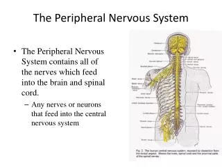
Histology of the Peripheral Nervous System. J. Matthew Velkey , Ph.D . matt.velkey@duke.edu 452A Davison, Duke South. special senses. Structural Organization of the Nervous System. inputs. outputs. Cardiac & smooth muscle & glands. Motor nerve. Skeletal muscle.

E N D
Histology of the Peripheral Nervous System J. Matthew Velkey, Ph.D. matt.velkey@duke.edu 452A Davison, Duke South
special senses Structural Organization of the Nervous System inputs outputs Cardiac & smooth muscle & glands Motor nerve Skeletal muscle
Functional Organization of the Nervous System • Somatic (conscious afferent* and efferent, voluntary motor control) • Autonomic(unconscious efferent, involuntary motor control of internal organs to maintain homeostasis) • Sympathetic– thoracolumbar division • Parasympathetic– craniosacral division * Somatic afferents = sensory fibers from skin, muscle, joints, tendons. Visceral afferents = sensory fibers from visceral organs; some result in conscious sensations, but others do not. However, they are not considered part of the autonomic nervous system, which is entirely efferent.
Spinal cord, DRG, Sympathetic and Parasympathetic Ganglia Netter pl. 154 IX parasympathetic X (vagus) Dorsal horns: Interneurons Ventral horns: Motor neurons Lateral horns: Sympathetic neurons Parasympathetic (S2-4) neurons DRG:Sensory (pseudounipolar) neurons Autonomic ganglia: Post ganglionic neurons with unmyelinated axons. Sympathetic: paravertebral ganglia Parasympathetic: In organs Somatic motor sensory Sympathetic: pre- & post-ganglionic fibers Pelvic splanchnic (parasympathetic)
Spinal cord and Dorsal Root Ganglion Dorsal median sulcus Dorsal horn (Lateral horn) if present Ventral horn Ventral median fissure Dorsal root ganglion (DRG)
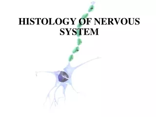
Cellular Components of the Nervous System Neurons Glia (support cells)
Generic neuron • Large cell body (aka soma or perikaryon) • Large, euchromaticnucleus (and usually a prominent nucleolus) • Extensive cytoplasmic extensions: • Dendrite(s): single or multiple extensions specialized for receiving input • Axon: single, large extension specialized for conveying output (in humans, can be up to 1.5m in length)
Motor neuron with Nissl bodies D NU N D NB NB AH V A D A-axon D-dendrite N-nucleus NB-Nissl body AH-axon hillock V-blood vessel NU-nucleolus
Synapses can form between many different parts of neurons and between a neuron and a non-neuronal cell, e.g., a muscle or a secretory cell. A single neuron can receive activating or inhibiting inputs from thousands of synaptic connections. Motor neuron cell body in the spinal cord
At a chemical synapse neurotransmitter release is triggered by the influx of Ca2+ and postsynaptic neurotransmitter receptors receive the signal.
An example of a synapse: The neuromuscular junction (motor endplate)
Conduction velocity in the axon is enhanced by myelinationaxons in the CNS are myelinated by oligodendrocytesaxons in the PNS are myelinated by Schwann cells
Myelination is a dynamic process, which involves the ensheathment of the the axon by the glial cell and subsequently the extrusion of cytoplasm from parts of the glial cell. Adhesive proteins on the cytoplasmic and the extracellular side of the plasma membrane contribute to a tight apposition of the lipid bilayers.
Myelinated Nerve Fiber The increased lipid content of the myelin sheath provides electrical insulation for the underlying axon. Myelin Sheath
Nodes of Ranvier are areas of the myelinated axon that are not covered by the myelin sheath. Ion channels are concentrated at the nodes of Ranvier and the myelin sheath acts as an electrical insulator. This allows for saltatory conductance of the action potential and increases the transmission speed of the nerve impulse.Depending on the diameter of the axon, myelination increases the action potential speed approximately 5 to 50fold (up to >110 m/sec).
Each Schwann cell myelinates a single internode Internode length can be up to 1.5 mm in the largest nerve fibers
One Schwann cell can ensheath multiple axons, but myelinates only one axon
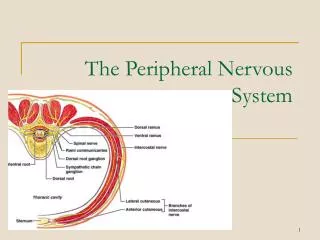
Connective tissue layers found in nerves:endoneurium surrounds axons,perineurium axon fascicles and epineurium the entire nerve
Connective tissue layers in a peripheral nerve. Tight junctions between perineurium cells form a important isolating barrier. Epineurium Perineurium
Sensory Ganglia • Two types: spinal (dorsal root) andcranial ganglia associated with spinal and cranial nerves, respectively • Contain large sensory neurons and abundant small glial cells, calledsatellite cells • Sensory neurons arepseudounipolar
Dorsal Root (Sensory) Ganglion Cells Dorsal root ganglion
Somatic sensory neurons have components in both CNS and PNS pseudounipolar sensory neuron in a dorsal root (spinal) ganglion Note that the spinal cord is part of the CNS and therefore does not contain Schwann cells, but rather oligodendrocytes. CNS PNS sensory input
Somatic motor neurons of the spinal cord also have components in the CNS and PNS, but they are multipolar Motor output: axon travels through peripheral nerve to reach target muscle
An example of sensory input:the muscle spindle Specialized skeletal muscle fibers enclosed within a spindle-shaped capsule. Depolarize in response to changes in muscle position, tension, and contraction velocity. Synapse with sensory nerve endings to convey input to CNS
The somatic nervous system in action:the spinal stretch reflex stretch receptor
Efferent autonomic pathways Sympathetic = thoracolumbar Parasympathetic = craniosacral
Pre-ganglionic motor neurons have components in the CNS and PNS and are also multipolar Visceral motor output to post ganglionic neuron
Sympathetic ganglion cells:multipolar neurons that reside entirely within the PNS in sympathetic chain ganglia and “pre-aortic” ganglia
Parasympathetic ganglion cells:multipolar neurons that also reside entirely within the PNSin the wall of the innervated organ (shown here in the seminal vesicle)
Objectives of PNS Histology: • Discuss the general division/differences between CNS and PNS • Appreciate the subdivision into somatic and autonomic nervous system • Learn about the cellular components and the structural attributes of neuronal cells • Discuss synaptic connections, using the motor end plate as an example • Study the formation of the axonal myelin ensheathment • Compare the histological features of myelinated and unmyelinated axons/nerves • Recognize nerves in histological sections • Identify the different connective tissue layers that are associated with nerves • Understand the different organizational plans that are adopted by neuronal cells • Identify and compare autonomic and sensory ganglia • Learn about the basic histological features of the spinal cord