Download

1 / 36
390 likes | 788 Views
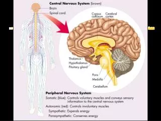
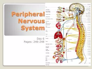
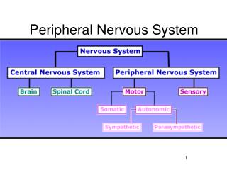
Peripheral Nervous System. Now that we’ve looked at spinal and cranial nerves , we can examine the divisions of the PNS. The PNS is broken down into a sensory and a motor division We’ll concentrate on the motor division which contains the 1. somatic nervous system and the

E N D
Now that we’ve looked at spinal and cranial nerves, we can examine the divisions of the PNS. • The PNS is broken down into a sensory and a motor division • We’ll concentrate on the motor division which contains the 1. somatic nervous system and the 2. autonomic nervous system.
Somatic vs. Autonomic • Voluntary • Skeletal muscle • Single efferent neuron • Axon terminals release acetylcholine • Always excitatory • Controlled by the cerebrum • Involuntary • Smooth, cardiac muscle; glands • Multiple efferent neurons • Axon terminals release acetylcholine or norepinephrine • Can be excitatory or inhibitory • Controlled by the homeostatic centers in the brain – pons, hypothalamus, medulla oblongata
Autonomic Nervous System • Motor subdivision of the PNS • Consists only of motor nerves • Also known as the involuntary nervous system • Regulates activities of cardiac and smooth muscles and glands • Two subdivisions • Sympathetic division • Parasympathetic division
ANS Structure • Both Sympathetic and Parasympathetic divisions have the same general structure. • Autonomic pathways always consist of 2 neurons in series. • They synapse in an autonomic ganglion • The 1st neuron in the autonomic pathway is the preganglionic neuron, • Cell body in CNS, myelinated, and projects to the autonomic ganglion. • While the 2nd neuron is the postganglionic neuron. • Cell body in autonomic ganglion, unmyelinated, and projects to the effector.
Anatomy of the Sympathetic Division • Originates from T1 through L2 • Ganglia are at the sympathetic trunk (near the spinal cord) • Short pre-ganglionic neuron and long post-ganglionic neuron transmit impulse from CNS to the effector • Norepinephrine and epinephrine are neurotransmitters to the effector organs
Anatomy of the Parasympathetic Division • Originates from the brain stem and S1 through S4 • Terminal ganglia are at the effector organ • Always uses acetylcholine as a neurotransmitter
Somatic and AutonomicNervous Systems Figure 7.27
Anatomy of the Autonomic Nervous System Figure 7.28
Sympathetic Pathways Figure 7.29
Autonomic Functioning • Sympathetic—“fight or flight” • Response to unusual stimulus • Takes over to increase activities • Remember as the “E” division • Exercise, excitement, emergency, and embarrassment • Parasympathetic—“housekeeping” activites • Conserves energy • Maintains daily necessary body functions • Remember as the “D” division • digestion, defecation, and diuresis
Antagonistic Control • Most internal organs are innervated by both branches of the ANS which exhibit antagonistic control A great example is heart rate. An increase in sympathetic stimulation causes HR to increase whereas an increase in parasympathetic stimulation causes HR to decrease
Exception to the dual innervation rule: • Sweat glands and blood vessel smooth muscle are only innervated by symp and rely strictly on up-down control. • Exception to the antagonism rule: • Symp and parasymp work cooperatively to achieve male sexual function. Parasymp is responsible for erection while symp is responsible to ejaculation. There’s similar ANS cooperation in the female sexual response.
Effects of the Sympathetic and Parasympathetic Divisions of the ANS Table 7.3 (1 of 2)
Effects of the Sympathetic and Parasympathetic Divisions of the ANS Table 7.3 (2 of 2)
Development Aspects of the Nervous System • No more neurons are formed after birth, but growth and maturation continues for several years • The brain reaches maximum weight as a young adult
Sympathetic vs. ParasympatheticStructural Differences: Sympathetic Parasympathetic
Sympathetic vs. ParasympatheticEffects: • In the following tables, note the effects of the sympathetic and parasympathetic nervous systems on various body organs.
Duration/Location of Parasympathetic Effects • Parasympathetic preganglionic neurons synapse on only a few postganglionic neurons. Would you expect parasympathetic activity to be widespread or local? • All parasympathetic fibers release ACh. • ACh is quickly broken down by what enzyme? What can you say about the duration of parasympathetic effects?
Why Is Sympathetic Activity Diffuse? • Preganglionic fibers have their somata in the lateral horns of the thoracic and lumbar spinal cord. • Preganglionic fibers leave the cord via the ventral root and enter a white ramus communicans to enter a chain ganglion – which is part of the sympathetic trunk. • Let’s look at a picture!
Once a preganglionic axon reaches the chain ganglion, it may: 1 2 …synapse with a ganglionic neuron w/i the same chain ganglion. …ascend or descend in the trunk to synapse within another chain ganglion. 3 …pass thru the chain ganglion and emerge from the chain w/o synapsing.
If the preganglionic axon synapses in a chain ganglion (routes 1 and 2)… It will enter the ventral or dorsal ramus of the adjoining spinal nerve via a gray ramus communicans. From here it may give branches to sweat glands, arrectorpili, and vascular smooth muscle – while it continues to its final destination which could be the iris muscles, the heart, or something else.
Preganglionic fibers that do not synapse in the trunk synapse with prevertebral ganglia located anterior to the vertebral column. • These are not arranged in a chain and occur only in the abdomen and the pelvis. • These are the splanchnic nerves. • Thoracic splanchnic nerves form a large plexus (abdominal aortic plexus) which yields multiple fibers that innervate visceral and vascular smooth muscle of the abdominal cavity. • Pelvic splanchnic nerves innervate the lower digestive organs (inferior large intestine) as well as urinary and reproductive structures.
Certain splanchnic nerves synapse on hormone-producing cells of the adrenal medulla – the interior of the adrenal glands which sit upon the kidneys. How does this contribute to the “diffuseness” of sympathetic activity?
How Does the Brain Control the ANS? • The hypothalamus is the Boss: • Its anterior and medial regions direct parasympathetic function while its posterior and lateral regions direct sympathetic function • These centers exert control directly and via nuclei in the reticular formation (e.g., the cardiovascular centers in the MO, respiratory centers in MO and pons, etc.) • The connection of the limbic system to the hypothalamus mediates our “flight or flight” response to emotional situations. • The relationship btwn the hypothalamus and the amygdala and periaquaductal gray matter allow us to respond to fear.