Download

1 / 98
1.11k likes | 1.96k Views
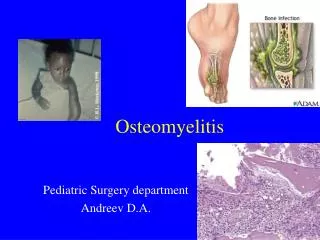
Osteomyelitis: Pathophysiology & Treatment Decisions. Clifford B. Jones, MD Associate Clinical Professor, Michigan State University Grand Rapids Orthopaedic Residency Program Orthopaedic Associates of Grand Rapids, Grand Rapids, MI Created March 2004; Revised February 2007.

E N D
Osteomyelitis:Pathophysiology & Treatment Decisions Clifford B. Jones, MD Associate Clinical Professor, Michigan State University Grand Rapids Orthopaedic Residency Program Orthopaedic Associates of Grand Rapids, Grand Rapids, MICreated March 2004; Revised February 2007
“One Should Especially Avoid Such Cases if One has a Respectable Excuse, for the Favorable Chances are Few and the Risks are Many….
….Besides, if a Man does not Reduce the Fracture, He will be Thought Unskillful. If He does Reduce It, He will bring the Patient Nearer to Death than Recovery.” Hippocratic Writings, New York, Pelican Books, 1978
Fracture Management Goals • Osseous Union • Restore Limb Function • Avoid Complications
Osteomyelitis Results in: • Reduction in limb function • Psychological & Social dysfunction • Increased cost
Hansen’s 7 DsConcerning Prolonged Orthopaedic Problems Despair Divorce Destitute Depression Delinquency Default Death Sigvard Ted Hansen, 1997
Introduction • 350,000 long bone fxs/yr • Infection risk varies: • Type I open – 10/1,000 infections • Type III open – up to 25%
Gustilo Open Fx ClassJBJS, 72A: 299-303, 1990 2% 7% 7% 10-50% 25-50%
Open Fractures Type II Type IIIA Type IIIB Type IIIB
Negative Biology of Open Fx Contamination Crushing Stripping Devascularization Comminution
Blood SupplyRhinelander, CORR, 1974 Normal - endosteal/medullary 2/3-3/4 internal external Fracture - periosteal/external majority internal external Periosteal Blood Supply Important
Initial Emergent Treatment dT Antibiotics, IV Reduce Stabilize Cover wound
Why infection risk high? Infection risk ≈ Fracture type (soft tissue) Open fx = Contamination (70% cx +) Open fx = Infected fx > 8 hours
Cost Analysis Infection • Increase cost 16-21%/pt • Increase hosp stay 36-50%/pt Total Cost $ 271 million/yr
Definition • Group of conditions • “…presence of bacteria & an inflammatory response causing progressive destruction of bone.” • Fears, RL, et al, 1998 • “…suppurative process in bone caused by a pyogenic organism” • Pelligrini, VD, et al, 1996
Why destruction of bone matrix? Proteolytic enzymes Hyperemia Osteoclasts
Classification • Waldvogel, 1971 • Classification based on pathogenesis • May, 1989 • 5 parts, post-traumatic tibial osteomyelitis • Cierny & Mader, 1985 • 4 factors affecting outcome • Host, site, extent of necrosis, degree of impairment
PathogenesisWaldvogel, 1971 • Hematogenous • Contiguous focus of infection • Direct inoculation
AnatomicClassification (Cierny-Mader) 1985 I: II: III: IV:
Classification Break-Down • Medullary Endosteal nidus, min soft tissue involvement, ? Sinus tract • Superficial Surface of bone, usu 2° to soft tissue defect • Localized Localized sequestra, usu sinus tract, Usu stables/p excision • Diffuse Permeative process, combination of I/II/III, Usu Unstable s/p excision
Physiologic Classification(Cierny-Mader, 1985) A-Host: Good immune system & delivery B-Host: Compromised host BL: locally compromised BS: systemically compromised BC: combined C-Host: Requires suppressive or no Tx Minimal disability Tx worse than dz, not a surgical candidate
Clinical Staging(Cierny-Mader, 1985) Anatomic Type +Clinical Stage Physiologic Class Example: IV BS tibial osteomyelitis = diffuse tibial lesion in a systemically compromised host
Types of Pathophysiology Acute/Hematogenous Chronic/Nonhematogenous
Acute/Hematogenous • Anatomy (Hobo) • Sharp twist in metaphyseal capillaries • Stasis (Trueta) • Decreased flow in capillaries & veins • Combination (Morrissy) • Trauma & Bacteria
Acute/HematogenousProgression of Dz • Cell death 2° to bacterial exotoxins • bacterial culture medium • worsens condition • Vascularity, leukocytosis, edema • Pressure w/in rigid osseous container • Pain, swelling, erythema • Potential for septic arthritis (knee, hip, shoulder)
Chronic/Nonhematogenous S. aureus ↑ Pseudomonas aureginosa ↑ Enterobacter > 30% Polymicrobial
Erythema Swelling Sinus Tract Drainage Limp Fluctuence None Pain Tenderness Fever HA Nausea/Vomiting Clinical Findings (varied)
Clinical Findings • Must have high index of suspicion • Inappropriate use of Abx – obscure Sx • Must obtain Dx quickly • If Tx started < 72°: • Decrease incidence of chronic osteomyelitis • Decrease destruction of bone
Laboratory Data Acute (Morrey, BF, OCNA, 1975) • WBC (25% of time) • Abnormal differential, Left Shift (65%) • Blood Cx – 50% positive Chronic • Mild anemia, WESR, C-reactive protein • Possible leukocytosis with L shift • Blood Cx – usually negative
Radiographs Early – usu negative Changes – delayed (10-21 days)
Radiographs Soft Tissue • Swelling, obscured soft tissue planes, haziness Osseous • Hyperemia, demineralization • Lysis (when > 40% resorbed) • Periosteal reaction • Sclerosis (late)
Radionucleotide Imaging 99M Tc 67Ga 111In WBC
99M Tc • Action • binds to hydroxyapetite crystals • Osteoblastic activity • Demineralized bone • Immature collagen
99M Tc • 3 Phase Bone Scan • Radionucleotide angiogram • Immediate post injection blood pool • Three hour: soft tissue, urinary excretion • Diagnosis • Cellulitis: Phases 1 &2, no change 3 • Osteomyelitis: Phases 1 & 2, focal 3 • Results: 94% sensitivity, 95% specificity • Rosenthal 1992, Schauwecker 1992
99M Tc: False Positive DM foot d/o Septic arthritis Inflammatory bone dz Adjacent to pressure sores
99M Tc 4 Phase Bone Scan • New development • Action: • Mature bone: uptake stops at 4 hr • Immature woven bone: cont’d uptake at 24 hr • Problem: needs f/u imaging at 24 hr (compliance) • Gupta 1988, Israel 1987, Schauwecker 1992
67Ga • Exudation of in vivo labeled serum protein • Transferrin, haptoglobin, albumin • Results • 81% sensitivity, 69% specificity • Schauwecker, 1992 • Combination with Tc • sensitivity, but specificity
111In WBC • Used in combination (Seabold, 1989) • In/Tc: 88% accurate • Ga/Tc: 39% accurate • Preparation problem • rad dose to spleen, 18-24hr delay • Spine (Whalen, Spine 1991) • 83% false negative use MRI
MRI No radiation Good soft tissue imaging Imaging: • T1 Dark • T2 Bright/Mixed
T1 bright T2 dark
T1 bright T2 dark
MRI • Acute: • marrow fat • granulation tissue H2O • Chronic: thickened cortex • Low signal on all scans • Cellulitis: no marrow changes
MRI ResultsSchauwecker, 1992 • Sensitivity 92-100% • Specificity 89-100% • Excellent for Spine (Modic, RCNA, 1986) • Sens 96%, Spec 92%, Accuracy 94% • Soft tissue extension • Sinus tract formation • Bright Tx from skin to bone
CT Imaging Image cortical and cancellous bone Evaluate osseous adequacy of debridement
Aspiration Biopsy Acute • Good, only 10-15% false negative Chronic • Sinus tract cx: 76% sens, 80% spec • 70% with S aureus & Enterococcus • 30% Pseudomonas • Does not determine correct Abx