Osteomyelitis
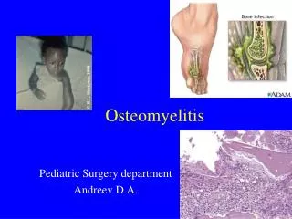
Osteomyelitis. Pediatric Surgery department Andreev D.A. How Common Is Osteomyelitis?.

Osteomyelitis
E N D
Presentation Transcript
Osteomyelitis Pediatric Surgery department Andreev D.A.
How Common Is Osteomyelitis? • Chronic osteomyelitis occurs in about 2 in 10,000 adults. Children have the acute form of the disease more often than adults do, at a rate of about 1 in 5,000. People who have diabetes, who have had a traumatic injury recently, or who use intravenous* drugs are at greatest risk for chronic infection.
Mortality/Morbidity • Mortality from osteomyelitis was 5-25% in the preantibiotic era. Presently, the mortality rate is approaching 0%. • Complications of osteomyelitis include • (1) septic arthritis, • (2) destruction of the adjacent soft tissues, • (3) malignant transformation (eg, Marjolin ulcer [squamous cell carcinoma], epidermoid carcinoma of the sinus tract), • (4) secondary amyloidoses, and • (5) pathologic fractures.
Cierny-Mader Staging System for Osteomyelitis • Anatomic type Stage 1: medullary osteomyelitis Stage 2: superficial osteomyelitis Stage 3: localized osteomyelitis Stage 4: diffuse osteomyelitis Physiologic class A host: healthy B host: Bs: systemic compromise Bl: local compromise Bls: local and systemic compromise C host: treatment worse than the disease Factors affecting immune surveillance, metabolism and local vascularity - Systemic factors (Bs): malnutrition, renal or hepatic failure, diabetes mellitus, chronic hypoxia, immune disease, extremes of age, immunosuppression or immune deficiency - Local factors (Bl): chronic lymphedema, venous stasis, major vessel compromise, arteritis, extensive scarring, radiation fibrosis, small-vessel disease, neuropathy, tobacco abuse • Adapted with permission from Cierny G, Mader JT, Pennick JJ. A clinical staging system for adult osteomyelitis. Contemp Orthop 1985;10:17-37
Organisms Commonly Isolated in Osteomyelitis Based on Patient Age • Infants (<1 year) Group B streptococci Staphylococcus aureus Escherichia coli Children (1 to 16 years)S. aureus Streptococcus pyogenes Haemophilus influenzae Adults (>16 years) Staphylococcus epidermidis S. aureus Pseudomonas aeruginosa Serratia marcescens E. coli • Adapted with permission from Dirschl DR, Almekinders LC. Osteomyelitis. Common causes and treatment recommendations. Drugs 1993;45:29-43.
The body is infected and the bacteria invade the blood through injured skin and mucous membranes, and the lymphoid throat ring. • Pyoderma of the skin, inflammation of the nasopharynx, and latent infections are of definite importance. • The umbilical wound is a frequent infection atrium in infants.
The anatomical age features of the structure and blood supply of the bones play a significant role in the development of osteomyelitis in children: • the richly developed network of blood vessels; • the autonomous supply of blood to the epiphysis, metaphysis, and diaphysis; • the presence of a great number of small vascular branchings stretching radially through the epiphyseal cartilage to the ossification nucleus. • The epiphyseal system of blood supply prevails in children under the age of 2 years, the metaphyseal system begins developing after this age. The epiphyseal and metaphyseal systems are isolated but there are anastomoses between them. The common vascular network forms only after ossification of the epiphysis.
Affection of the epiphyseal zone is characteristic of children under the age of 2-3 years. • With age, when the system of blood supply to the metaphysis begins developing intensively, it is the metaphysis that predominantly becomes affected.
Pain • which is a consequence of hypertension in the marrow cavity, is indirect proof of this interpretation of the circulatory disorders in the bone. Intraosseous pressure in acute osteomyelitis reaches 300-500 mm water (normal value in healthy children, 60-100 mm water).
If the osteomyelitic process is not recognized • when it is still in the stage of inflammation within the boundaries of the bone-marrow cavity, then beginning from the 4th or 5th day of the disease the pus spreads along the bony haversian and Volkmann's canals under the periosteum and gradually separates it. • Later (the 8th to 10th day and later) pus and the products of disintegration continueseparating the periosteum, then the pus breaks through into the soft tissues and forms intermuscular and subcutaneous phlegmons.
Clinical picture • The toxic (adynamic) form follows an extremely violent course with signs of endotoxic shock. A state of collapse is observed as a rule, with loss of consciousness, delirium, high body temperature (up to 40-41 °C), and sometimes with convulsions and vomiting. • Dyspnoea is found but without any clear clinical picture of pneumonia. • The cardiovascular abnormalities include disorders of central and peripheral circulation, reduced arterial pressure, with the development within a short time of cardiac insufficiency and signs of myocarditis. • Punctate extravasations are often seen on the skin. • The tongue is dry and with a brownish coating. The abdomen is usually distended and tender in the upper parts. The liver is enlarged.
Septicopyaemic form The onset of the disease is also acute: • body temperature rises to a high level (39-40°C), • signs of toxicosis increase, and the activity of vital organs and systems is disturbed. • Confused consciousness, delirium, and euphoria are sometimes encountered. • Pain is experienced in the affected limb from the first days of the disease and becomes very intense due to the development of intraosseous hypertension. • Septic complications caused by the spread of the purulent foci to various organs (the lungs, heart, and kidneys, as well as to the other bones) often occur.
Thelocalized form • characterized by the predominance of local signs of purulent inflammation over the general clinical manifestations of the disease
The main constant local signs of osteomyelitis • sharp local tenderness to palpation and particularly to percussion over the site of the lesion. • Oedema and tenderness extend also to the adjoining areas. • Such signs as hyperaemia of the skin and fluctuation in the region of the lesion are very late signs and are evidence of neglected osteomyelitis
The main constant local signs of osteomyelitis • Considerable diagnostic difficulties arise in osteomyelitis of bones forming the hip joint. The local signs are indistinct on the first days of the disease due to the powerful muscular casing in this region. • On careful inspection it can be seen that the lower limb is slightly flexed at the hip joint; abduction and mild external rotation. • Movements at the hip joint are painful. The joint itself and the overlying skin are oedematous.
Findings in infants include the following: • Failure to thrive • Drowsiness but irritability • Minimal constitutional symptoms • Effusions into neighboring joints (60%)
Findings in older children include the following: • History of preceding minor trauma to the involved limb and/or recent infection, eg, upper respiratory tract or skin infection • Bone pain • Malaise, irritability, and anorexia • Fever • Reluctance to use the limb • Localized swelling, redness, and warmth • Tenderness to finger pressure at a particular point • Pain on moving an adjacent joint • Regional lymphadenopathy
The X-ray signs of acute haematogenic osteomyelitis are manifested no earlier than on the 14th-21st day of the disease.
The X-ray signs • Reduced density of the bone shadow and blurring of its contours are usually found, osteoporosis in the region corresponding to the zone of the inflammation can also be detected. The spongy substance of the bone produces a macromacular pattern due to resorption of the bony trabeculae and merging of the intertrabecular spaces as the result of intensified resorption.
Nuclear medicine • Nuclear medicine bone scans are a highly sensitive (>90%) modality in the diagnosis of osteomyelitis. This procedure is done in 3 parts. Technetium Tc 99m is used to create images to determine areas of infection and bone remodeling dependent on local blood flow. The sensitivity of bone scans is often helpful when the exact site and extent of the infection is not known.
MRI • MRI if available is another useful modality for imaging acute osteomyelitis. Findings on MRI accurately illustrate the extent and structure of the area involved in the pathologic process. Sensitivity has been reported to be 88-100%, with a specificity of 75-100%. Fat-suppression sequences allow for better detection of bone marrow edema; however, infection and inflammation cannot be differentiated. MRI may be the imaging modality of choice in infections involving the spine, pelvis, or limbs because of its ability to provide fine details of the osseous changes and soft-tissue extension in these areas.
Limitations of Techniques: • MRI has limited availability and is relatively expensive. MRI is also contraindicated in patients with certain implant devices and metallic clips, and it is not tolerated by all patients because of claustrophobia or morbid obesity. In addition, young children may requiring sedation, Good MRI require patient cooperation because patient motion can degrade the images. • CT is quick and inexpensive, but exposes the patient to ionizing radiation. The risk of a reaction to radio-iodinated contrast material is low, though the detection of bone destruction or a paraspinal mass does not require the use of contrast material. • Although radionuclide studies are sensitive, they can be time-consuming, and they have lower spatial resolution. The incidence of false-negative scans is low in neonates and in elderly patients with osteomyelitis.
Diagnosis of osteomyelitis • Diagnostic puncture of the bone with subsequent cytological examination of the aspirated material should be carried out more extensively in questionable cases. • Measurement of intraosseous pressure is very important in establishing the early diagnosis of acute haematogenic osteomyelitis. The discovery of intraosseous hypertension confirms the diagnosis even in the absence of pus under the periosteum and in the marrow cavity.
Diagnosis of osteomyelitis • Blood tests show leukocytosis (up to 30 000-40 000 per mm3) with a shift of the differential count to the left and toxic neutrophil granulation. The ESR is markedly increased (up to 60 mm/hour) and remains high for a long time. • Marked changes are found in the blood serum protein spectrum. These are dysproteinaemia, an increase in the globulin fractions, and the development of hypoalbuminaemia. Anaemia caused by bone marrow inhibition by the prolonged effect of toxins develops in a persisting and severe disease. • Disorders of the blood coagulation system are also found (the fibrinogen concentration and the fibrinolytic activity increase, the recalcification time and the coagulation time become shorter, the prothrombin index increases).
differential diagnosis • articular form of rheumatism, • phlegmon, • tuberculosis of the bones, • and injury.
Rheumatism is characterized by shifting pains in the joints and typical changes in the heart confirmed by electrocardiography. Careful inspection and palpation of the involved region reveals that in rheumatism, in contrast to osteomyelitis, tenderness and swelling are mainly localized over the joint and not over the bone. Improvement of the local process with the prescription of salicylates is an important factor
Tuberculosis of the bones • Though experiencing pain in the limb, the child still uses it. • Alexandrov's sign (thickening of the skin fold on the involved limb) and muscle atrophy are found. The radiograph demonstrates osteoporosis (the "melting sugar" symptom,) and an indistinct periosteaLreaction. This reaction, however, maybe clearly pronounced in mixed infection and in accompanying ordinary flora. The so-called acute forms of osteoarticular tuberculosis are actually cases of delayed diagnosis made when pus has already penetrated the joint. In addition to the X-ray picture, identification of the specific causative agent in material aspirated from the joint helps in establishing the correct diagnosis.
Abscesses of the psoas muscle • The classic presentation includes fever, back pain and a limp. Common clinical signs include a positive psoas sign (pain when the hip is passively extended or actively flexed against resistance), which is attributed to inflammation causing spasm of the psoas muscle, and femoral neuropathy, which includes a limp or a flexion deformity of the involved hip.
Abscesses of the psoas muscle • CT scanning is an accurate, rapid and noninvasive method for diagnosing psoas abscess and delineating its cause. • Extraperitoneal surgical drainage has been the standard method of treatment; however, image-guided percutaneous drainage has become an effective alternative.
Treatment • In view of the fact that most severe forms of osteomyelitis are consequent upon intraosseous hypertension, early surgical intervention, osteoperfora-tion, acquires primary importance. An incision, no less than 10-15 cm in length, is made in the soft tissues overlying the lesion and the periosteum is cut longitudinally. Two or three perforating openings 3-5 mm in diameter are made at the junction with the healthy bone. Pus is usually discharged under pressure in such cases, while in a disease of a long duration the contents of the marrow cavity may be seropurulent for two or three days. The marrow cavity is irrigated with 1 : 5000 nifrofurazone solution and antibiotics through the perforation in the bone.
Metaepiphyseal osteomyelitis • is mostly encountered among infants, predominantly among the newborn. By the haematogenic route the infection (usually staphylococcus) enters the bone metaphysis and the inflammatory process develops here. Due to the peculiar blood supply of the metaepiphyseal junction in very young children, however, the inflammation spreads to the growth zone and epiphysis located in the joint. As a result, the main clinical symptoms are caused by the developing acute arthritis.
Clinical picture • Metaepiphyseal osteomyelitis sets in acutely as a rule with a rise of body temperature, debility, refusal of food, reluctance to move the involved limb which the child holds in a forced position. • Examination reveals swelling over the zone of affection, deformity of the'adjoining joint, increase of local temperature. Hyperaemia appears later. Palpation and passive movement of the limb cause sharp pain. The "pseudoparesis" symptom (the hand or foot of the involved limb hangs and movements in it are sharply limited). The local form of osteomyelitis may be complicated by phlegmon of the soft tissues around the joint.
The X-ray signs • are demonstrated earlier in metaepiphyseal osteomyelitis than in the other forms. Some characteristic signs can be detected as early as the 8th-10th day: thickening of soft tissues on the affected side, widening of the X-ray joint space, a fine periosteal reaction . Foci of destruction in the metaphysis are demonstrated on the radiographs only on the 3rd week after the onset of the disease, whereas the degree of destruction of the bone epiphysis