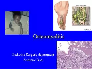
Osteomyelitis
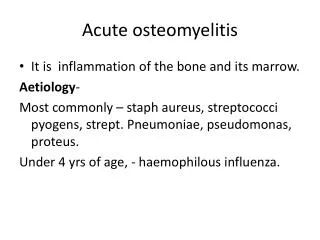
Osteomyelitis. Always a Diagnostic Puzzle. schreibman.info. Osteomyelitis: Put the Pieces Together. HISTORY Clinical Surgical. RADIOGRAPHS Recent . MRI Active. CT Chronic. Osteomyelitis: Topics. Definitions Active Chronic Mechanisms Hematogenous Direct spread Imaging


Osteomyelitis
E N D
Presentation Transcript
Osteomyelitis Always a Diagnostic Puzzle schreibman.info
Osteomyelitis: Put the Pieces Together • HISTORY • Clinical • Surgical • RADIOGRAPHS • Recent • MRI • Active • CT • Chronic
Osteomyelitis: Topics • Definitions • Active • Chronic • Mechanisms • Hematogenous • Direct spread • Imaging • Radiographs • CT • MRI Bone Model Cortex Marrow
Osteomyelitis: Definitions “Osteomyelitis” • comes from Greek: • osteon = bone • myelos = marrow • itis = inflammation • “Inflammation of bone marrow” • Infection of bone marrow • High Sensitivity • Low Specificity • Marrow inflammation from infection looks like inflammation from any other cause • MRI • Marrow
Osteomyelitis: Definitions • Active Osteomyelitis • vs • Chronic Osteomyelitis
Osteomyelitis: Definitions • Active Osteomyelitis • “Aggressive” • Resembles Tumor • Cortex Destruction • Periosteal Reaction
Active Osteomyelitis 16yoM distal fibula pain 3w after inversion injury • “Aggressive” • Cortex Destruction • Periosteal Reaction • HISTORY Clinical Followup
Osteomyelitis: Definitions • Chronic Osteomyelitis • “Non-Aggressive” • Resembles Callus 3 Characteristics: • Involucrum: “wrap” • Thick periosteum around infected bone • Sequestrum: “set apart” • Piece of dead, infected, bone • Cloaca: “sewer” • Opening in cortex throughwhich pus can escape RADIOGRAPHS Active ≠ Chronic
Active vs Chronic Osteomyelitis RADIOGRAPHS Active ≠ Chronic Active Osteomyelitis Chronic Osteomyelitis
Active Osteomyelitis 16yoM distal fibula pain 3w after inversion injury “Aggressive” • Cortex Destruction • Periosteal Reaction Active Osteomyelitis
Chronic Osteomyelitis 19yoM fibula pain2.5years later… RADIOGRAPHS Active ≠ Chronic Chronic Osteomyelitis 2.5 years
Chronic Osteomyelitis 19yoM fibula pain2.5years later… CT Tibia Involucrum Chronic Osteomyelitis Fibula Sequestrum Cloaca
Chronic Osteomyelitis 42yoM Diabetic • Involucrum Developing 6 weeks later 10 more weeks
Chronic Osteomyelitis 27yoM s/p removal Rt Femoral Rod 27yoM s/p removal Rt Femoral Rod • Involucrum CT Scout
Chronic Osteomyelitis 27yoM s/p removal Rt Femoral Rod 27yoM s/p removal Rt Femoral Rod • Involucrum • Sequestrum Axial Slice CT Scout Coronal Reformat
Chronic Osteomyelitis 27yoM s/p removal Rt Femoral Rod 27yoM s/p removal Rt Femoral Rod • Involucrum • Sequestrum • Cloaca Axial Slice CT Scout Oblique Coronal
Osteomyelitis: Mechanisms • Direct Spread adjacent tissues • Most common cause • Decubitus ulcer • Septic arthritis PUS
Decubitus UlcerIschium 52yoM quadriplegic T1 Ischium Ischium T1
Osteomyelitis: Mechanisms • Direct Spread adjacent tissues • Most common cause • Decubitus ulcer • Septic arthritis • Puncture into bone • Stepped on nail • External fixator • Ring sequestrum
Ring Sequestrum • Chronic Osteomyelitis • Involucrum • Sequestrum • Cloaca • Poor Union RADIOGRAPHS
Osteomyelitis: Mechanisms • Direct Spread adjacent tissues • Most common cause • Decubitus ulcer • Septic arthritis • Puncture into bone • Stepped on nail • External fixator • Ring sequestrum • Hematogenous • Site related to patient age
Hematogenous Osteomyelitis • Site related to patient age Epiphysis Physis Metaphysis Arteriole Venule Infection occurs at end of Infection occurs at metaphysis of Septic Emboli Diaphysis Blood Supply Mature Bone Immature Bone
Hematogenous Osteomyelitis 1yoM strep pneumonia
Hematogenous Osteomyelitis 1yoM strep pneumonia 3 months later
Osteomyelitis: Imaging • Many Imaging Options: • Radiographs • CT • MR • US • Nuc Med • What to order when?
Osteomyelitis: What to Order When • Radiographs ………… ALWAYS! • May show evidence of active infection • Bone destruction, periosteal reaction • May show evidence of chronic infection • Involucrum • Screen for metal • Orthopedic hardware, foreign bodies • Unexpected findings • Fractures, • Delineate current anatomy • Surgical resections, • RADIOGRAPHS NEED TO BE RECENT vs gas in soft tissues neuropathic deformity
Need for Recent Radiographs Example • 66yoM h/o Diabetes • Presents in Sept swollen foot • MR is requested to “r/o Osteo” • Are there radiographs? • Yes …3 months ago • Repeat radiographs obtained now, prior to MR, reveal… Neuropathic destruction of the Lisfranc joint Normal Lisfranc joint June September
Osteomyelitis: What to Order When • Radiographs ………… ALWAYS! • CT……………………. Chronic Cases • CT best for calcified structures • Involucrum • Sequestrum • Cloaca • CT of the extremities is insensitive for: • Bone marrow pathology • Soft tissue pathology
Osteomyelitis: What to Order When • Radiographs ………… ALWAYS! • CT……………………. Chronic Cases • MRI..…………………. Active Cases • Shows extent of soft tissue edema • Excellent for demonstrating abscesses and other drainable fluid collections • Sensitive for bone marrow pathology • Can be overly sensitive at expense of specificity • Infected bone marrow resembles marrow edema due to other causes
Osteomyelitis: MR Imaging Bone Model Marrow Cortex X-rays T1 T2
Osteomyelitis: MR Imaging Bone Model Marrow Marrow Marrow Cortex Cortex Cortex Surrounding Surrounding Tissues (fat) Tissues (fat) X-rays T1 T2
Osteomyelitis: MR Imaging Bone Model Marrow Marrow Marrow Cortex Cortex Cortex Surrounding Surrounding Tissues (fat) Tissues (fat) X-rays T1 T2fs
Osteomyelitis: MR Imaging • Path=Fluid • T1=Dark • T2=Bright • T1fs+Gd • Enhancement • Inflamed Uniform • Abscess Wall • Cyst Not (STIR) T1 T2fs
Osteomyelitis: MR Imaging • Enhancement • Inflamed Uniform • Abscess Wall • Cyst Not (STIR) T1fs T1 T2fs +Gd
Osteomyelitis: MR Imaging • Detection of the non-enhancing pus pocket (abscess) is crucial • Presence of soft tissue abscess proves the edema in underlying bone marrow is osteomyelitis. • Site for aspiration for culture. • If IV Gd doesn’t get into abscess, IV antibiotics won’t get in either, abscess may require drainage.
Osteomyelitis: MR Imaging 63yoM Diabetic with heel ulcer Intact cortex T1 • Enhancing cellulitis • No non-enhancingabscess pocket Minimal Marrow Edema Arterial Ca++ T1fs IVGd IR
Osteomyelitis: MR Imaging 63yoM Diabetic 2 weeks later… 2 weeks earlier Cortical destruction Intact cortex • Non-enhancingabscess pocket • More marrow edema • More tissue edema T1fs IVGd IR
Osteomyelitis: MR Imaging 63yoM Diabetic 2 weeks later… 2 weeks earlier Cortical destruction Intact cortex Marrow edema Abscess Pocket T1fs IVGd T1 IR
Decubitus UlcerIschium 52yoM quadriplegic Abscess! T1fs +Gd T1 Abscess? T2fs
Osteomyelitis: MR Imaging 1yoF Swollen left lower leg R L Periosteal Reaction Metaphyseal lucency
Osteomyelitis: MR Imaging 1yoF Swollen left lower leg Brodie Abscess Periosteal Reaction Non-enhancing abscess • Intra-osseous Metaphyseal T1fs IVGd T1 T2fs
Osteomyelitis: What to Order When • Radiographs ………… ALWAYS! • CT……………………. Chronic Cases • MRI..…………………. Active Cases • US……….…………… Fluid/Abscess • US guided aspiration for culture • Cannot assess bone involvement • Nuc Med.……………. Problem Cases • Where MR specificity is decreased • Neuropathic feet • Infected hardware
Infection around metal: MRI T2fs T1fs IVGd We can see soft tissues around bone Enhancing granulation tissue (phlegmon?) We can’t see the marrow within bone Cannot evaluate for “osteomyelitis” T,K 21yoM
Infection around metal: Nuc Med • Requires 2 Radiopharmaceuticals • Tc-Bone Scan(Active bone metabolism) • In-WBC Scan (Areas of WBC accumulation) 1)BS: Sen/Spec 2)WBC:Spec/Sen
Infection around metal: Nuc Med Removed Tibia Plate Tc-Bone Scan In-WBC Scan Femur Plate Femur Femur Tibia Tibia Placed Antibiotic PMM-Beads Tibia Plate S,B 31yoM
Charcot (Neuropathic) Foot T1 T1fs +IV Gd T2fs Abscess Tc99m MDP In111 WBC Infection P,K 65yoF
Osteomyelitis: Put the Pieces Together • HISTORY • Clinical • Surgical • RADIOGRAPHS • Recent • MRI • Active • CT • Chronic