Download
1 / 46
490 likes | 620 Views
Explore the intricate workings of the neuromuscular junction and muscle stimulation process, including details on motor units, polarization, depolarization, propagation of action potential, sliding filament mechanism, and energy sources. Gain insights into fine and coarse muscle control mechanisms.

E N D
Motor Unit • one motor neuron • all the skeletal muscles it stimulates
Fine Muscle Control • few muscle fibers stimulated by one motor neuron • single motor neuron may supply very few fibers (eye) • Result: - finer control of muscle fibers
Coarse Muscle Control • many muscle fibers stimulated by one motor neuron • single motor neuron may supply many fibers (large muscle) • Result: - less control of muscle fibers
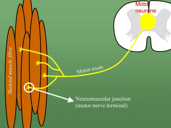
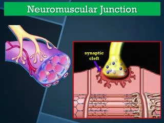
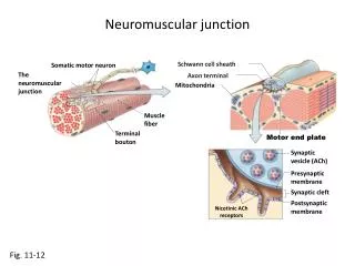
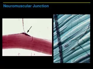
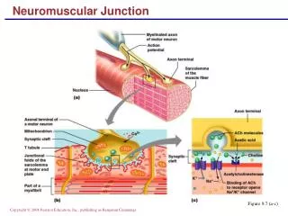
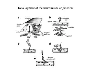
Neuromuscular Junction • contact or junction between motor neuron and a skeletal muscle - thread-like extensions of neuron branch into many axonal terminals - each branch forms a junction with sarcolemma (one muscle fiber)
Nerve Endings • individual branches of axon near muscle fiber loose myelin sheath • divide into several bulb-shaped structures (synaptic end bulb) - bulbs contain neurotransmitter acetylcholine (ACh)
Nerve Endings • extremely close to muscle but never touch • space is called synaptic cleft - filled with interstitial fluid
Motor End Plate • portion of muscle fiber membrane adjacent to synaptic end bulb of motor neuron • contains receptors for acetylcholine
Polarization • muscle fiber relaxed (resting sarcolemma) • outside sarcolemma + charge (predominant extracellular ion is Na+) • inside sarcolemma - charge (predominant intracellular ion is K+) • sarcolemma is relatively impermeable to both ions
Depolarization(generation of the action potential) • stimulation of sarcolemma by motor nerve • patch of sarcolemma becomes permeable to sodium ions (sodium gates open) • + sodium ions rush into cell • inside sarcolemma + charge • outside sarcolemma - charge • this rush upsets electrical currents causing action potential
Propagation of the Action Potential • + charge inside sarcolemma changes permeability of adjacent patches on sarcolemma • depolarization is repeated - therefore action potential spreads along entire length of sarcolemma
Repolarization • events occur in reverse • sarcolemma permeability changes • Na+ gates close • K+gates open allowing diffusion of K+ ions out of cell • activation of sodium-potassium pump restores ionic resting state concentrations
Repolarization (cont.) • occurs in same direction as depolarization • must occur before muscle can be stimulated again
Muscle Stimulation • muscle fibers are stimulated by motor neurons • impulse arrives at axon terminal of motor neuron
Muscle Stimulation (cont.) • impulse depolarizes plasma membrane opening voltage-sensitive calcium channels (Ca+2) • calcium ions diffuse from extracellular fluid into the axon terminal - triggers release of acetylcholine from synaptic end bulb
Muscle Stimulation • ACh diffuses across synaptic cleft • ACh interacts with receptors in the motor end plate of the muscle fiber, thus altering its permeability to sodium ions (Na+) • sodium ions diffuse from extracelluar fluid into muscle fiber, producing local depolarization called end-plate potential
Muscle Stimulation(power stroke) • end-plate potential generates flow of ions or current to bring adjacent sarcolemma to threshold • current spreads in both directions triggering action potentials • action potentialinitiate wave of contraction by way of transverse tubules
Muscle Stimulation (power stroke) (cont.) • action potential triggers release of Ca+2 from sarcoplasmic reticulum • Ca+2 ions bind to troponin molecules on the thin filaments • tropomyosin moves, uncovering cross-bridge binding sites on actin • binding of actin and myosin causes ATP to split releasing energy for the power stroke
Muscle Stimulation (power stroke) (cont.) • rotational movement of a myosin cross-bridge • one power stroke of a cross-bridge results in a small movement of the thin filament • each cross-bridge produces many cycles of movement during a single twitch contraction
Muscle Stimulation (power stroke) (cont.) • acetylcholine is quickly decomposed by cholinesterase • its decomposition prevents generation of further end-plate potentials
Sliding Filament Mechanism • during muscle contraction, neither the thick nor the thin filaments decrease in length • the actin (thin) filaments slide like pistons inward among the myosin (thick) filaments
Sliding Filament Mechanism (cont.) • in the resting state, the ends of the actin barely overlap the myosin • during contraction, these ends overlap considerably while the two Z membranes approach the ends of the myosin filaments
Myosin has globular bridges Ca+2 ions help cross bridges react with actin Actin ADP molecules on surface act as sites for linkages with cross bridges
Sources of Energy • phosphate system • glycogen-lactic acid system • aerobic system
Phosphate System • ATP and creatine phosphate • together they provide energy for muscles to contract maximally for approximately 15 seconds • this system is used for short bursts of energy
Energy Source for Muscle Contraction • immediate source is ATP(adenosine triphosphate) • supplied by mitochondria near myofibrils • enzyme ATPase splits a phosphate group from ATP, forming ADP (adenosine diphosphate) and P (phosphate group) • energy released when P is split from molecule of ATP activates myosin cross-bridges
Energy Source for Muscle Contraction • very little ATP present in muscle fibers • if exercise is to continue for more than a few seconds, additional ATP must be produced
Energy Source for Muscle Contraction • primary energy source available to regenerate ATP from ADP and phosphate is creatine phosphate • contains high-energy phosphate bonds • cannot directly supply energy to a cell • 3-5 times more abundant in muscle fibers than ATP
Creatine Phosphate • stores energy released from mitochondria • when sufficient ATP is present, creatine phosphokinase (enzyme) promotes synthesis of creatine phosphate • energy is stored in its phosphate bonds • when ATP is being decomposed, energy from CP is transferred to ADP and then quickly converted back to ATP
Glycogen-Lactic Acid System • with continued activity, muscles require energy after the supply of creatine phosphate is depleted • glucose must be catabolized to generate ATP
Glycogen-Lactic Acid System • glucose passes into contracted muscles via blood (facilitated diffusion) • glucose is also produced by glycolysis (breakdown of glycogen in muscles)
Glycolysis • series of ten reactions • splits glucose into two molecules of pyruvic acid and two molecules of ATP • anerobic process (no oxygen)
Glycogen-Lactic Acid System • pyruvic acid formed by glycolysis enters mitochondria - its oxidation produces large quantities of ATP from ADP • some activities do not supply enough O2 to completely break down pyruvic acid • pyruvic acid is then converted to lactic acid
Glycogen-Lactic Acid System (cont.) • most lactic acid diffuses from skeletal muscles into the blood • heart muscle fibers, kidney cells and liver cells use lactic acid to produce ATP • liver cells can convert lactic acid back to glucose • some lactic acid is accumulated in blood and muscles
Glycogen-Lactic Acid System (cont.) • can provide energy for about 30-40 seconds of maximal muscle activity, e.g., a 50 meter swimming race
Aerobic System • reactions that require oxygen carried by the blood • oxygen is bonded to molecules of hemoglobin
Cellular Respiration • when energy is exhausted, muscles become dependant upon cellular respiration of glucose as a source of energy for synthesis of ATP • muscle activity longer than 30 seconds requires an aerobic process
Aerobic System • conversion of pyruvic acid into CO2, H20, and ATP • yields 36 molecules of ATP from each glucose molecule • provides energy for muscular activity lasting longer than 30 seconds
Recovery Oxygen Consumption (oxygen debt) • elevated oxygen use after exercise • above resting oxygen consumption • elevated oxygen necessary to restore metabolic conditions to resting state
Recovery Oxygen Consumption • converts lactic acid back into pyruvic acid • reestablishes glycogen stores in muscle and liver cells • resynthesizes creatine phosphate and ATP • replaces O2 removed from myoglobin
Recovery Oxygen Consumption(cont.) • ATP production for metabolic reactions (increased rate due to increased body temperature) • ATP production for continued elevated activity of cardiac and skeletal muscles • ATP production needed for an increased rate of tissue repair
Muscle Fatigue(inability of a muscle to contract) • Condition may result from: - insufficient O2 delivered to muscle cells - depletion of glycogen stored in muscle cells - buildup of lactic acid in body fluids - insufficient acetylcholine