Download
1 / 40
1.08k likes | 5.62k Views
Neuromuscular Junction Disorders. Nuha Alkhawajah MD. Definition of NMJ Disorders. Disorders affecting the junction between the presynaptic nerve terminal and the postsynaptic muscle membrane Pure motor syndromes Preferentially affect proximal, bulbar, or extraocular muscles.

E N D
Neuromuscular Junction Disorders NuhaAlkhawajah MD
Definition of NMJ Disorders • Disorders affecting the junction between the presynaptic nerve terminal and the postsynaptic muscle membrane • Pure motor syndromes • Preferentially affect proximal, bulbar, or extraocularmuscles
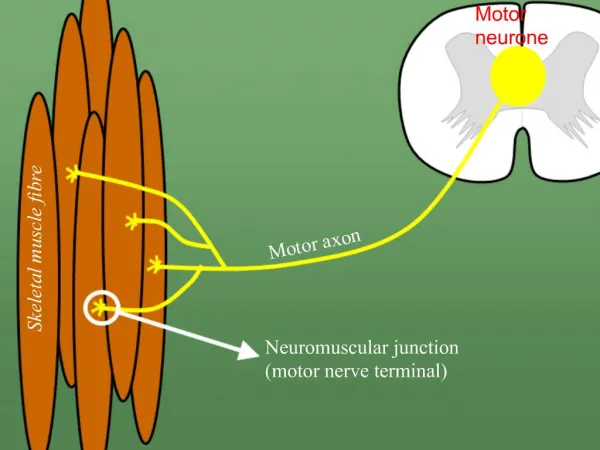
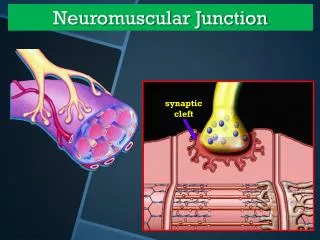
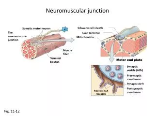
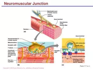
Physiology of NMJ Transmission • chemical neurotransmitter at the NMJ is acetylcholine(ACH) • ACH is stored in vesicles in the presynaptic terminal in discrete units known as quanta • Each quantum contains 10,000 molecules of ACH • The quanta are located in three separate stores. The primary, or immediately available store, secondary and a tertiary far from the NMJ in the axon and cell body.
Physiology of NMJ Transmission • Action potential invades and depolarizes the presynaptic junction • VGCCs are activated, allowing an influx of calcium • Release of ACH • ACH binds to ACHRs on the postsynaptic muscle membrane • This opens sodium channels • Leading to local depolarization, the endplate potential (EPP)
Physiology of NMJ Transmission • The size of the EPP is proportional to the amount of ACH released • A threshold needs to be reached for the EPP to be produced and muscle fiber action potential to be generated • In normal circumstances, the EPP always rises above threshold, resulting in a muscle fiber action potential • In the synaptic cleft, ACH is broken down by the enzyme acetylcholinesterase
Myasthenia Gravis (MG) • The most common disorder of neuromuscular transmission • Caused by an immunoglobulin G (IgG)-directed attack on the NMJ nicotinic ACH receptor • A post-synaptic NMJ disorder. • Hallmark of the disorder is a fluctuatingfatigable weakness
Myasthenia Gravis (MG) • Classification • According to onset • Congenital • Acquired • According to clinical presentation: • Ocular • Generalized
Epidemiology of MG • Prevalence is 200 per million • Bimodal distribution: • Early peak: 2nd and 3rd decades (female predominance) • Late peak: 6th to 8thdecade (male predominance) • Neonatal MG: a transient form, due to trans-placental passage of maternal antibodies • Association with other autoimmune diseases as, autoimmune thyroid disease, SLE, and rheumatoid arthritis,neuromyelitisoptica
Pathogenesis of MG • Autoantibodies against the AChR • Decrease in the number of active acetylcholine as a consequence of AChR antibody binding • Destruction of receptors occurs via a complement-mediated process • Destruction of the post-synaptic folds • Associated with thymus pathology.
Pathogenesis of MG • 60-70% of AChR ab positive patients have thymic hyperplasia and 10-12% have thymoma • Produces AChR subunits that triggers the immune response
Clinical features of MG worsening contractile force, not tiredness • Fluctuating, intermittent symptoms sometimes with periods of spontaneous improvement • Appearing with repetitive activity and worsening as day progresses • Muscle fatigue and weakness • Noabnormality of mental state, sensoryor autonomic function • Characteristically affects the extra-ocular, bulbar or proximal limb muscles
Types of Presentation in MG • Ocular presentation 50% • Bulbar presentation 15% • Limb weakness (<5 percent) • Isolated neck (uncommon) • Isolated respiratory (rare)
Ocular-onset MG • the most common • eventually 90% • 15% continue to have isolated ocular symptoms • Ptosis (droopy eyelids) • Extraocularweakness frequently begins asymmetrically • Mimics 3rd , 4th , and 6thnerve palsies and, rarely INO • Unlike true 3rd nerve palsies MG never affects pupillary function
The normal eyelid and palpebral fissure • normal eyelid crease is 6 to 7 mm away from the eyelid margin in adults. • upper eyelid covers top 1 mm of the cornea • normal PF measures 9 to 12 mm • distance from a central pupillary light reflex to upper eyelid margin is called the margin reflex distance, normally this measures 4 to 5 mm
Bulbar-onset MG • Bulbar muscles weakness is the next most common • Dysphagia • Fatigability and weakness of mastication, with the inability to keep the jaw closed after chewing. • Dysarthria: nasal speech, slurred and hypophonic • Nasal regurgitation
Facial muscles involvement • Facial muscles are frequently involved • Patient appear expressionless • "myasthenic sneer” on attempting to smile where the mid-lip rises but the outer corners of the mouth fail to move
Limb involvement in MG • limbs weakness, usually symmetric and proximal • wrist and finger extensors and foot dorsiflexors are often involved • Rare patients present with an isolated limb weakness and never develop eye movement or bulbar muscle weakness
Respiratory Involvement in MG • Difficulty breathing, SOB • Obstructive sleep apnea • Difficulty sleeping on flat bed
Diagnosing MG • Bed side tests: • Tensilon test: injection of edrophonium (acetylcholinesteraseinhibitor) in patients with ptosis or ophthalmoparesis looking for improvement • Ice pack test
Diagnosing MG • Serologic testing: • Antiacetylcholine receptor antibodies (AChR-Ab): • 80-90% of generalized MG • 50% of ocular MG • Anti Muscle-specific kinase antibodies (MuSK-Ab): • 38-50% of generalized MG who are AChR-Ab –ve • much lower frequency of thymic pathology • More common in females • Usually present with severe oculobulbar weakness along or neck, shoulder, and respiratory weakness
Diagnosing MG • Electrophysiological studies: • repetitivenerve stimulation studies • single-fiber EMG the most sensitive test • CT scan of the chest. Why?
Prognosis • Early, the symptoms are often transient, with hours, days, or even weeks free of symptoms • Symptoms typically worsen and are more persistent months later. • Maximum weakness is reached within two years in 82 percent of patients
Prognosis • An active phase with fluctuations and most severe symptoms in the 1stfive to seven years. Most myasthenic crises occur in this early period. • More stable second phase, symptoms are stable but persist. They may worsen in the setting of infection, medication taper, or other perturbations. • Followed by 3rdphase, in which remission may occur
Treatment of MG • Crisis: IVIG or Plasma exchange • Symptomatic treatment: cholinestrase inhibitor “Pyridostigmine” • Chronic immunomodulatory and immunosuppressive treatment:steroids, azathioprine, cellcept…. • Thymectomy
Myasthenic Crisis • life-threatening condition • Definition: weakness from acquired MG that is severe enough to necessitate intubation • due to weakness of respiratory muscles. • Severe oropharyngeal muscle weakness often accompanies the respiratory muscle weakness, or may be the predominant feature
Cont. • Triggered by infections or certain medications. • A list of medications that affect the NMJ transmission should be given to MG patients to avoid or to use with caution.
Lambert Eaton Myasthenic Syndrome • Presynaptic NMJ disorder • Middle age to old people • 50% of cases are associated with malignancy (especially lung cancer) • Fluctuating proximal weakness • Associated with P/Q type voltage gated Ca channels antibodies
Botulism • Presynaptic NMJ disorder • Caused by toxin produced by Clostridium Botulinum • Inhibits the release of Ach from the NMJ, sympathetic and parasympathetic ganglia • Food borne or wound related • Descending weakness and autonomic disturbance