Neuromuscular junction
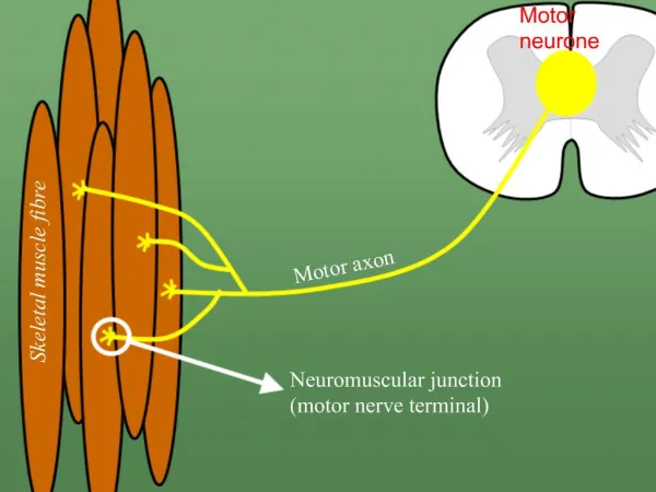
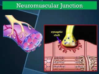
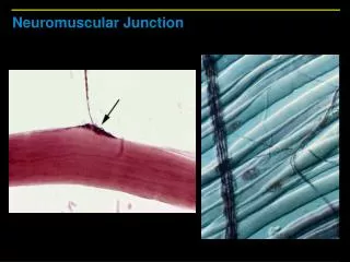
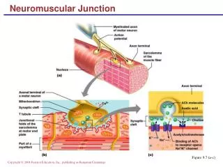
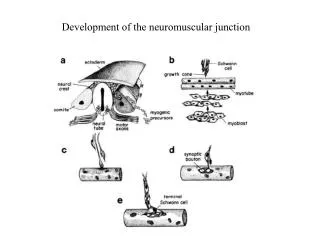
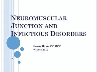
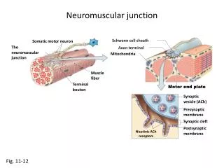
Neuromuscular junction. Schwann cell sheath. Somatic motor neuron . The neuromuscular junction. Axon terminal. Mitochondria. Muscle fiber. Terminal bouton. Motor end plate. Synaptic vesicle (ACh). Presynaptic membrane. Synaptic cleft. Postsynaptic membrane. Nicotinic ACh

Neuromuscular junction
E N D
Presentation Transcript
Neuromuscular junction Schwann cell sheath Somatic motor neuron The neuromuscular junction Axon terminal Mitochondria Muscle fiber Terminal bouton Motor end plate Synaptic vesicle (ACh) Presynaptic membrane Synaptic cleft Postsynaptic membrane Nicotinic ACh receptors Fig. 11-12
Neuromuscular junction Somatic motor neuron Axon terminal Ca2+ Ca2+ Action potential ACh Acetyl + choline Voltage-gated Ca2+ channel Skeletal muscle fiber AChE Motor end plate Nicotinic receptor Open channel Closed channel K+ Na+ ACh K+ Na+ Fig. 11-13
T-tubules and SR T-tubule brings actionpotentials into interiorof muscle fiber. Thin filament Thick filament Sarcolemma Sarcoplasmic reticulumstores Ca2+ Fig. 12-4
Excitation-contraction coupling Axon terminal ofsomatic motor neuron Muscle fiber ACh Na+ Motor end plate RyR T-tubule Sarcoplasmicreticulum Ca2+ DHP Z disk Troponin Actin Tropomyosin M line Myosin head Myosin thick filament Ca2+ released Myosin thick filament Fig. 12-11
Troponin and tropomyosin Thin filaments Troponin Nebulin Tropomyosin G-actin molecule Actin chain Fig. 12-3
Cross-bridge cycle 1 Cytosolic Ca2+ Troponin G-Actin 3 Tropomyosin shifts,exposing bindingsite on actin 2 TN TN 5 Myosin head Actinmoves Tropomyosinblocks bindingsite on actin ADP ADP 4 Pi Power stroke Pi Relaxed state. Myosin head cocked. Initiation of contraction Fig. 12-9
Types of Muscle Fibers Fig. 12-15
Motor Units One muscle may havemany motor units ofdifferent fiber types. SPINAL CORD Neuron 1 Neuron 2 Neuron 3 Motornerve KEY Motor unit 1 Muscle fibers Motor unit 2 Motor unit 3 Fig. 12-15
Basic circulatory system Fig. 9-2c, p.360
Extrinsic Muscle Pump Extrinsic muscle Valve Blood
Blood vessel structure Fig. 15-2
Mammalian circulation Fig. 14-1
External anatomy of the heart Aorta Pulmonaryartery Superiorvena cava Auricle ofleft atrium Rightatrium Coronaryarteryand vein Rightventricle Leftventricle Fig. 14-7
Internal anatomy of the heart Pulmonarysemilunar valve Aorta Rightpulmonaryarteries Left pulmonaryarteries Superiorvena cava Left pulmonaryveins Right atrium Left atrium Cusp of the AV(bicuspid) valve Cusp of a right AV(tricuspid) valve Chordae tendineae Papillary muscles Left ventricle Right ventricle Inferiorvena cava Descendingaorta Fig. 14-7
Cardiac muscle structure Intercalateddisks Myocardial musclecell Fig. 14-7