Download

1 / 46
460 likes | 735 Views
Testicular tumors and STDs. Dr. Basu MD. Our topic. Classification of testicular tumor. Seminoma Embryonal carcinoma Yolk sac tumor Choriocarcinoma Teratoma Diagnosis of these tumors. What you should know about a Testicular tumor. Age Gross and microscopy Markers

E N D
Testicular tumors and STDs Dr. Basu MD
Our topic • Classification of testicular tumor. • Seminoma • Embryonal carcinoma • Yolk sac tumor • Choriocarcinoma • Teratoma • Diagnosis of these tumors
What you should know about a Testicular tumor • Age • Gross and microscopy • Markers • Clinical Presentation
Classification of testicular tumor. • Tumor arising from the Germ cells • Tumor arising from Leydig cells( produce endocrine abnormality). • Tumor arising from Sertoli cells.
Tumor arising from the Germ cells • Tumors with one histological pattern • Seminoma • Embryonal carcinoma • Yolk sac tumor • Choriocarcinoma • Teratoma • Tumor with more than one histological pattern • Miscellaneous
Seminoma [Classic] • Most common types of testicular neoplasm. • Age : 15 to 34 years • Note: • Some Seminoma may contain trophoblastic content. • In these type of Seminoma Beta-HCG will be mildly elevated.
Variant of Seminoma Variant : Spermatocytic Seminoma • In this case metastasis is rare, common in old people. • Three types of cell are seen • large multinucleated cells, • medium size cells and • small cells that reminiscent of spermatocytes
Seminoma Gross Features : large, soft, homogenous, grey-white
Seminoma : Microscopy • Seminoma cells ([ PAS positive] : Large cells with distinct border , round nuclei and prominent nucleoli. • Lymphocytes, plasma cell in stroma.
Seminoma counterpart in Ovary DYSGERMINOMA
Embryonal carcinoma Age : 20-30 years. Features : • Often multiple metastasis is present at the time of diagnosis. • Often it contain other foci of Yolk sac tumor, teratoma and Chorio-carcinoma. • So both AFP and beta hcG will be elevated ( non specific)
Embryonal carcinoma Features : Red to tan to brown areas, including prominent hemorrhage and necrosis.
Embryonal carcinoma and Teratoma [ Teratocarcinoma] Features : Chondroid white areas (teratoma) in a Embryonal carcinoma.
Teratoma in testis • Age = all ages • Almost always malignant ( unlike ovary – where it is usually benign)
Yolk sac tumor [ endodermal sinus tumor] • Age : 3 years • Histology : Presence of Schiller –Duvall body ( glomeruloid body) • Specific Marker = AFP
Schillar Duval body – glomeruloid structurein yolk sac tumor ; locate it
Choriocarcinoma • Age = 20 -30 • Pure Chorio carcinoma is rare in testis. • It is always mixed with Teratoma, or other tumor even with Seminoma. • Histology : Malignant cyto and syncytiotrophoblast without villous formation. • Specific Marker = beta hcG
Mixed tumor • Add………….
Leydig cell tumor : Clinical features Small( 1-3 cm), nodular, circumscribed tumor, yellowish in colour Bilateral gyenecomastia and testicular enlargement force the patient to seek medical assistance.
Quiz : name the markers AFP hcG AFP hcG AFP and hcG
Testicular tumor; clinical features • Painless swelling • Seminoma usually confined to testis. • Other non-seminomatous tumor widely metastasize . • Metastasis occur by both hematgenous and lymphatic route.
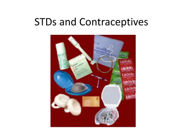
Secondary syphilis Strongly Positive both • Anti treponomal antibody test and • Nontrepomomal test
Positive anti treponomal antibody test. Negative – Nontrepomomal antibody test Syphilis
Condylomata acuminata ( HPV infection type 6,11) : Genital Warts
Gonorrhea ; clinical features • Male : Epididymitis, may involve prostate. • Female : salpingitis, infertility • Infants ( during delivery) : Purulent infection of the eye : Ophthalmia neonatorum).
Lymphogranuloma Venereum, LGV lymphadenopathy. Mixed Granulomatous and neutrophilic inflammation.
Diagnosis - LGV • Demonstration of organism in Biopsy section / exudates- in active lesion. • ELISA performed on serum.
“Soft chancre” –Chancroid in Hemophilus ducreyiinfection. Ulcer contain yellowish exudates.
Syphilis ( Primary- Painless clear base ulcer, no exudates) ; hard chancre
Syphilis ( secondary – maculopapular rash) Histology shows plasma cells and lymphocytes
Syphilis - Secondary :: Condylomata lata -This broad base, elevated lesion seen in the moist areas.
Cause of false positive VDRL test • SLE • Lepromatous leprosy • Pregnancy • Antiphospholipid syndrome
Granuloma Inguinale ; ulcerated papular lesion Calymmatobacterium donovani
Genital herpes simplex: Painful erythematous vesicles Etiology : HSV type 2 and 1