Download

1 / 42
680 likes | 2.01k Views
Testicular tumors. B.Vijay Anand, SRMC, Chennai. . Incidence . Testicular tumors are rare. 1 – 2 % of all malignant tumors. Most common malignancy in men in the 15 to 35 year age group. Benign lesions represent a greater percentage of cases in children than in adults. Age - 3 peaks

E N D
Testicular tumors B.Vijay Anand, SRMC, Chennai.
Incidence • Testicular tumors are rare. • 1 – 2 % of all malignant tumors. • Most common malignancy in men in the 15 to 35 year age group. • Benign lesions represent a greater percentage of cases in children than in adults.
Age - 3 peaks 2 – 4 yrs 20 – 40 yrs above 60 yrs • Testicular cancer is one of the few neoplasms associated with accurate serum markers. • Most curable solid neoplasms and serves as a paradigm for the multimodal treatment of malignancies.
Etiology • Cryptorchidism • Intersex disorder • Testicular atrophy • Trauma- prompts medical evaluation • Chromosomal abnormalities - loss of chromosome 11, 13, 18, abnormal chromosome 12p. • Sex hormone fluctuations, estrogen administration during pregnancy
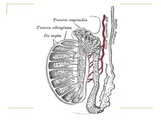
CROSS SECTION OF TESTIS Testis Stroma Seminiferous Tubules (200 to 350 tubules) Interstitial Cells Supporting Spermatogonia Leydig or (Androgen) Sertoli Cell
CLASSIFICATION I. Primary Neoplasms of Testis. A. Germ Cell Tumor. B. Non-Germ Cell Tumor . II. Secondary Neoplasms. III. Paratesticular Tumors.
Germ cell tumors 1. Seminomas - 40% (a) Classic Typical Seminoma (b) Anaplastic Seminoma (c) Spermatocytic Seminoma 2. Embryonal Carcinoma - 20 - 25% 3. Teratoma - 25 - 35% (a) Mature (b) Immature 4. Choriocarcinoma - 1% 5. Yolk Sac Tumour
Classification of germcell tumor (GCT) • GCTs arise from pluripotential cells, so a variety of elements may habitate in primary tumor • More than half of GCTs contain more than one cell type and are therefore known as mixed GCTs
Sex cord/ gonadal stromal tumors ( 5 to 10% ) 1. Specialized gonadal stromal tumor (a) Leydig cell tumor (b) sertoli cell tumor 2. Gonadoblastoma 3. Miscellaneous Neoplasms (a) Carcinoid tumor (b) Tumors of ovarian epithelial sub types
II. SECONDARY NEOPLASMS OF TESTIS A. Reticuloendothelial Neoplasms B. Metastases III. PARATESTICULAR NEOPLASMS A. Adenomatoid B. Cystadenoma of Epididymis C. Desmoplastic small round cell tumor D. Mesothelioma E. Melanotic neuroectodermal
Carcinoma insitu {CIS} • Pre invasive precusor of all GCT, except spermatocytic seminoma • Incidence of CIS in the male population is 0.8%. • Testicular CIS develops from fetal gonocytes & is characterized histologically by seminiferous tubules containing only Sertoli cells and malignant germ cells.
Patients at risk of CIS • History of testicular carcinoma (5% to 6%), • Extra gonadalGCT (40%), • Cryptorchidism (3%), • Contralateral testis with unilateral testis cancer (5% to 6%), • Somatosexual ambiguity (25% to 100%) • Atrophic testis 30 % • Infertility (0.4% to 1.1%) • TESTICULAR BIOPSY gold standard for diagnoses of CIS
Lymphatic drainage • The primary drainage of the right testis is within the interaortocaval region. • Left testis drainage , the para-aortic region in the compartment bounded by the left ureter, the left renal vein, the aorta, and the origin of the inferior mesenteric artery. • Cross over from right to left is possible.
Lymphatic drainage • Lymphatics of the epididymis drain into the external iliac chain. • Inguinal node metastasis may result from scrotal involvement by the primary tumor, prior inguinal or scrotal surgery, or retrograde lymphatic spread secondary to massive retroperitoneal lymph node deposits. • Testicular cancer spreads in a predictable and stepwise fashion, except choriocarcinoma. .
Clinical features • Painless Swelling of One testis • Dull Ache or Heaviness in Lower Abdomen • 10% - Acute Scrotal Pain • 10% - Present with Metatstasis - Neck Mass / Cough / Anorexia / Vomiting / Back Ache/ Lower limb swelling • 5% - Gynecomastia • Rarely - Infertility
Physical Examination • Examine contralateral normal testis. • Firm to hard fixed area within tunica albugenia is suspicious • Seminoma expand within the testis as a painless, rubbery enlargement. • Embryonal carcinoma or teratocarcinoma may produce an irregular, rather than discrete mass.
Differential Diagnosis • Testicular torsion • Epididymitis, or epididymo-orchitis • Hydrocele, • Hernia, • Hematoma, • Spermatocele, • Syphilitic gumma .
DICTUM FOR ANY SOLID SCROTAL SWELLINGS • All patients with a solid, Firm Intratesticular Mass that cannot be Transilluminated should be regarded as Malignant unless otherwise proved.
Scrotal ultrasound • Ultrasonography of the scrotum is a rapid, reliable technique to exclude hydrocele or epididymitis. • Ultrasonography of the scrotum is basically an extension of the physical examination. • Hypoechoic area within the tunica albuginea is markedly suspicious for testicular cancer.
Tumor markers TWO MAIN CLASSES • Onco-fetal Substances : AFP & HCG • Cellular Enzymes : LDH & PLAP AFP - Trophoblastic Cells HCG - Syncytiotrophoblastic Cells ( PLAP- placental alkaline phosphatase, & LDH lactic acid dehydrogenase)
AFP –( Alfafetoprotein) NORMAL VALUE: Below 16 ngm / ml HALF LIFE OF AFP – 5 and 7 days Raised AFP : • Pure embryonal carcinoma • Teratocarcinoma • Yolk sac Tumor • Combined tumors, • AFP not raised in pure choriocarcinoma , & in pure seminoma
HCG – ( Human Chorionic Gonadotropin) Has and polypeptide chain NORMAL VALUE: < 1 ng / ml HALF LIFE of HCG: 24 to 36 hours RAISED HCG - 100 % - Choriocarcinoma 60% - Embryonal carcinoma 55% - Teratocarcinoma 25% - Yolk Cell Tumour 7% - Seminomas
ROLE OF TUMOUR MARKERS • Helps in Diagnosis - 80 to 85% of Testicular Tumours have Positive Markers • Most of Non-Seminomas have raised markers • Only 10 to 15% Non-Seminomas have normal marker level • After Orchidectomy if Markers Elevated means Residual Disease . • Elevation of Markers after Lymphadenectomy means a STAGE III Disease
ROLE OF TUMOUR MARKERS • Degree of Marker Elevation Appears to be Directly Proportional to Tumor Burden • Markers indicate Histology of Tumor: If AFP elevated in Seminoma - Means Tumor has Non-Seminomatous elements • Negative Tumor Markers becoming positive on follow up usually indicates - Recurrence of Tumor • Markers become Positive earlier than X-Ray studies
Imaging studies • Chest X ray • CECT abdomen – retroperitoneal nodes • PET- No apparent advantage over CT • MRI - No apparent advantage over CT
Large left para aortic nodal mass due to GST causing hydronephrosis
Tumor staging • Primary Tumor (T)pTX - Primary tumor cannot be assessed (if no radical orchiectomy has been performed, TX is used) • pT0 - No evidence of primary tumor (e.g., histologic scar in testis) • pTis - Intratubular germ cell neoplasia (carcinoma in situ) • pT1 - Tumor limited to the testis and epididymis and no vascular/lymphatic invasion • pT2 - Tumor limited to the testis and epididymis with vascular/lymphatic invasion or tumor extending through the tunica albuginea with involvement of tunica vaginalis • pT3 - Tumor invades the spermatic cord with or without vascular/lymphatic invasion • pT4 - Tumor invades the scrotum with or without vascular/lymphatic invasion
Regional Lymph Nodes • Clinical NX - Regional lymph nodes cannot be assessed • N0 - No regional lymph node metastasis • N1 - Lymph node mass 2 cm or less in greatest dimension or multiple lymph node masses, none more than 2 cm in greatest dimension • N2 - Lymph node mass, more than 2 cm but not more than 5 cm in greatest dimension, or multiple lymph node masses, any one mass greater than 2 cm but not more than 5 cm in greatest dimension • N3 - Lymph node mass more than 5 cm in greatest dimension
Pathologic node staging • pN0 - No evidence of tumor in lymph nodes • pN1 - Lymph node mass, 2 cm or less in greatest dimension and ≤6 nodes positive, none >2 cm in greatest dimension • pN2 - Lymph node mass, more than 2 cm but not more than 5 cm in greatest dimension; more than 5 nodes positive, none >5 cm; evidence of extranodal extension of tumor • pN3 - Lymph node mass more than 5 cm in greatest dimension.
Distant metastasis • M0 - No evidence of distant metastases • M1 - Nonregional nodal or pulmonary metastases • M2 - Nonpulmonary visceral masses
PRINCIPLES OF TREATMENT • Treatment should be aimed at one stage above the clinical stage • Seminomas - Radio-Sensitive. Treat with Radiotherapy. • Non-Seminomas are Radio-Resistant and best treated by Surgery • Advanced Disease or Metastasis - Responds well to Chemotherapy
PRINCIPLES OF TREATMENT • Radical INGUINAL ORCHIDECTOMY is Standard first line of therapy • Lymphatic spread initially goes to RETRO-PERITONEAL NODES • Early hematogenous spread RARE • Bulky Retroperitoneal Tumours or Metastatic Tumors Initially “DOWN-STAGED” with CHEMOTHERAPY
PRINCIPLES OF TREATMENT • Transscrotal biopsy is to be condemned. • The inguinal approach permits early control of the vascular and lymphatic supply as well as en-bloc removal of the testis with all its tunicae. • Frozen section in case of dilemma.
CHEMOTHERAPY Chemotherapy Toxicity BEP - Bleomycin Pulmonary fibrosis Etoposide (VP-16) Myelosuppression Alopecia Renal insufficiency (mild) Secondary leukemia Cis-platin Renal insufficiency Nausea, vomiting Neuropathy
Lymph Nodes Dissection For Right & Left Sided Testicular Tumours
CONCLUSION • Improved Overall Survival of Testicular Tumour due to Better Understanding of the Disease, Tumour Markers and Cis-platinum based Chemotherapy. • Current Emphasis is on Diminishing overall Morbidity of Various Treatment Modalities .