Download
1 / 29
290 likes | 376 Views
This comprehensive study guide explores the lymphatic system, blood components, immune response, and related topics. Learn about lymphatic vessels, blood cells, leukemia, and blood typing. Discover the functions of lymphatic organs and the flow of lymph in the body. Gain insights on lymph nodes, white blood cell identification, and hematocrit interpretation. Study the physiology of lymph nodes, leukocytes, and blood circulation. Understand the significance of differential white blood cell counts and blood typing in healthcare settings. Enhance your knowledge of immune response mechanisms and hematological disorders. This guide provides essential information for students and professionals in the medical field.

E N D
Lab 5 –Blood, Lymphatics and the Immune Response Gilbert Pitts, Ph.D., Joseph Schiller, Ph.D., James F. Thompson, Ph.D.
Objectives • Examine: • Lymph node slide • Lymphatic vessels on charts and models • Blood slides • Differentiate red blood cells, platelets, neutrophils, lymphocytes, monocytes, eosinophils, and basophils • Perform 2 differential WBC counts • Identify leukemia • Identify sickle cell anemia • Understand blood typing (ABO and Rh systems) • Calculate and interpret hematocrit/packed cell volume
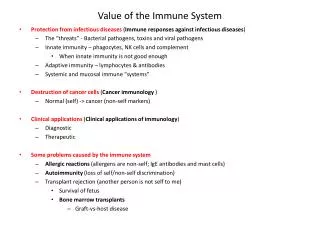
The Lymphatic System • Basic organization • Lymph fluid in lymph vessels • Lymphatic organs (red bone marrow, thymus, spleen, lymph nodes, tonsils) with smaller collections of lymphatic tissue in other organs • Functions • Return interstitial fluid and proteins to the blood • Transport dietary fats to adipose tissue • Protect against cancer & infection • Lymph Flow from smallest to largest: • Capillaries vessels trunks ducts • Lymph vessels anastomose and supply and drain lymph nodes along their course
Lymph Flow Follows Venous Circulation • Right head, arm, and thorax drained by the right lymphatic duct into right subclavian vein • Left head, arm, thorax, most of the abdominal cavity and both legs drained by the thoracic duct into the left subclavian vein R L
The Lymph Node • Connective tissue capsule with trabeculaeextending from cortex to medulla • Stroma – the supportive connective tissue network of reticular fibers and fibroblasts trabeculae capsule
Circulation in the Lymph Nodes • Lymph enters via a number of afferent lymphatic vessels • It then enters a large subcapsular sinus and travels into a number of smaller sinuses • It meanders through these sinuses and exits the node at the hilus via efferent vessels • The node acts as a “settling tank,” because there are fewer efferent vessels, lymph stagnates somewhat in the node • This allows lymphocytes and macrophages time to carry out their protective functions Only lymph nodes filter lymph! Metastasis: cancer cells from a tumor break away and flow with the lymph until trapped in the lymph nodes
Lymph Node Parenchyma • Cortex - filled with lymphocytes and macrophages in follicles • Outer edge of follicle contains more T cells • Inner germinal center is the site of B-cell proliferation • Medulla - medullary cords of lymphocytes, macrophages, plasma cells (activated B cells) Cortex Medulla
Lymph Node Micrograph Medulla Cortex
Lymph Node Germinal Centers germinal centers
The Formed Elements of the Blood: • Leukocytes = White Blood Cells • Granular leukocytes (granulocytes) • neutrophils • eosinophils • basophils • Agranular leukocytes (agranulocytes) • lymphocytes - T cells, B cells • monocytes tissue macrophages
Eosinophil 2-4% Neutrophil 60-70% Basophil 0.5-1% Granular Leukocytes
Lymphocyte 20-25% Monocyte 3-8% Agranular Leukocytes
Leukocyte Life Span and Number • 5,000 - 10,000 WBC’s/mm3 blood • RBC/WBC ratio 700/1 • Differential WBC count (a standard clinical lab report) • Neutrophils 60-70% • Lymphocytes 20-25% • Monocytes 3-8% • Eosinophils 2-4% • Basophils 0.5-1% • Abnormal proportions are correlated with different types of disease processes
Eosinophil 2-4% Lymphocyte 20-25% Monocyte 3-8% Neutrophil 60-70% Basophil 0.5-1% Differential WBC Count
Leukocyte Identification Agranular Granular All have many large granules in cytoplasm & multilobed nuclei Dark Hidden nuc. Small Spherical nucleus Basophil Lymphocyte Red gran. Large 2+ lobes Eosinophil Faint gran. Monocyte no large granules in cytoplasm Neutrophil
Composition of Blood • Blood sample separates into 2 parts • plasma - straw colored liquid on top • ~55% of the volume • formed elements • ~45% of the volume • red blood cells • buffy coat: white blood cells and platelets
Hematocrit (Hct) • Packed Cell Volume is the % of the blood which is RBC’s • Males: 40-54% (47%) • Females: 38-46% (42%) • Hct indicates the status of RBC production, the state of hydration, or various disease states
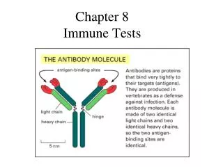
Blood Typing • Antigen – any substance which provokes specific immune responses • Antigenic determinants • Antigen parts which trigger the specific immune response • An antigen may be an entire microbe or only small structures such as subregions of large molecules • RBC antigens (agglutinogens) are membrane glycoproteins Most “antigens” are complex and express multiple types of antigenic determinants.
ABO Blood Types • 2 glycoprotein agglutinogens, A & B • One gene from each parent, A, B or O • 6 combinations - AA, AB, AO, BB, BO, OO (no agglutinogens)
ABO Blood Types • Agglutinins • Naturally occurring antibodies produced in response to the agglutinogens not present in your blood • React in antigen-antibody response to blood not of your type • blood type AB = universal recipients • blood type O = universal donors
Rh System • Rh typing - Rhesus monkey • Those expressing Rh antigens are Rh+ • Those without Rh agglutinogens are Rh- • Normally, blood does not contain Rh agglutinins • Immune system only makes agglutinins in response to specific exposure to Rh antigens • Rh sensitivity does not occur until second transfusion • Hemolytic disease of the newborn (erythroblastosis fetalis) • many “blue babies” prior to WWII
Please Clean Up Your Work Area • Place only lancet and capillary tubes in designated sharps containers • Place all other blood contaminated materials (gloves, alcohol wipes, paper towels, etc.) in the large red biohazard bucket at the front of the room • Place all other discards in regular trash receptacles • Disinfect your work area with the spray solution after you have concluded your blood work.
Homework • Complete and turn in the questions on pages 5-13 to 5-15 • Complete Assignment 5 on MasteringAandP.