BLOOD
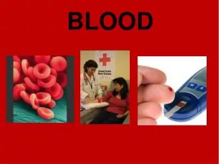
BLOOD. Blood transports oxygen maintains homeostasis fights infection prevents blood loss maintains body temperature. Hematophobia = fear of blood. Blood and Blood Cells. Blood is a type of CONNECTIVE TISSUE It has two basic components: Plasma (water, proteins, amino acids..etc) = 55%

BLOOD
E N D
Presentation Transcript
Blood • transports oxygen • maintains homeostasis • fights infection • prevents blood loss • maintains body temperature Hematophobia= fear of blood
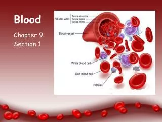
Blood and Blood Cells Blood is a type of CONNECTIVE TISSUE It has two basic components: Plasma (water, proteins, amino acids..etc) = 55% CELLS (rbc, wbc, platelets) = 45%
Plasma Proteins Albumins – blood pressure Globulins– transport lipids and antibodies for immunity Fibrinogen – important for blood clotting MAJOR EVENT IN BLOOD CLOTTING = Fibrinogen converted to FIBRIN
Hematocrit - volume of blood cells in a sample, should be 45%. The remaining fluid is plasma (55%). To determine the percentages, blood is placed in a centrifuge
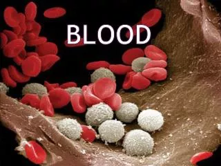
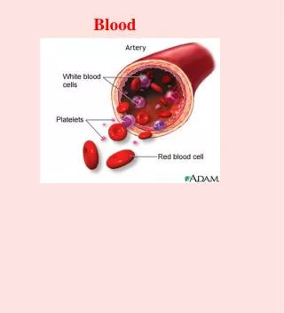
Three Types of Blood Cells red blood cells (erythrocytes)white blood cells (leukocytes)platelets (thrombocytes)
Main Functions of RBCs Transports oxygen, picks up carbon dioxide HEMOGLOBIN - molecule that combines with O2 IRON is critical to synthesize hemoglobin
Erythrocytes (RBCs) Figure 17.3
WHITE BLOOD CELLS(Leukocytes)) General function is to protect the body against disease There are FIVE different kinds of WBCs Granulocytes (granular cytoplasm) Neutrophils, Eosinophils, Basophils Agranulocytes (lacking granular cytoplasm) Monocytes, Lymphocytes
Neutrophil(nucleus has several lobes) Active phagocytes 60% of WBC Present in the pus of wounds
Eosinophil Mainly attack parasites 2% WBC
Basophil Produces Heparin and Histamines Important in Inflammatory Reaction 1% WBC
Monocyte(larger cell, horseshoe shaped nucleus) Become macro-phages
Lymphocyte(nucleus is dark and takes up almost whole cell; almost no cytoplasm seen) Defense against invaders Yield Antibodies 30% WBC
Platelets (thrombocytes) Blood clots and vessel repair
HEMOSTASIS The process of stopping bleeding Involves the coagulation and clotting of the blood to seal the site of damage
THREE EVENTS IN HEMOSTASIS 1. Blood Vessel Spasm Seratonin = vasoconstrictor 2. Platelet plug formation 3. Blood coagulation conversion of fibrinogen to fibrin
Hemostasis Blood Clot Formation Animated(Video) 2D animation Medivisual
COAGULATION - the thickening of blood to form a clot (hematoma)
THROMBUS – blood clot (abnormal) EMBOLUS – when the clot moves to another place.
Stages of Differentiation of Blood Cells Figure 17.9
Blood Type is Controlled by 3 Alleles 4 Possible Blood Types Alleles: A, B, O A & B are codominant O is recessive
Blood that has antibodies on it that is not recognized by the body will be attacked by your immune system
ABO Blood Groups Table 17.4
Rh Factor A person can either be Rh + or Rh – (positive is dominant)
Rh Factor and Pregnancy *Problem: When a fetus is Rh+ and the mother is Rh-, this can cause the mother’s immune system to attack the fetus. There are drugs that will suppress this reaction.
The right side receives oxygen-poor blood from the body and tissues and then pumps it to the lungs to pick up oxygen and dispel carbon dioxide Its left side receives oxygenated blood returning from the lungs and pumps this blood throughout the body to supply oxygen and nutrients to the body tissues The heart=a muscular double pump with 2 functions Overview
Cone shaped muscle Four chambers Two atria, two ventricles Double pump – the ventricles Two circulations Systemic circuit: blood vessels that transport blood to and from all the body tissues Pulmonary circuit: blood vessels that carry blood to and from the lungs simplified…
It weighs about 1 lb. Heart’s position in thorax Feel your heart beat at apex (this is of a person lying down)
CXR(chest x ray) Normal male
Chest x rays Lateral (male) Normal female
Starting from the outside… Pericardium(see next slide) Without most of pericardial layers
Muscle of the heart with inner and outer membrane coverings Muscle of heart = “myocardium” The layers from out to in: Epicardium = visceral layer of serous pericardium Myocardium = the muscle Endocardium lining the chambers Layers of the heart wall
Two atria (divided by interatrical septum) Right atrium Left atrium Two ventricles (divided by interventricular septum) Right ventricle Left ventricle Chambers of the heartsides are labeled in reference to the patient facing you --------------------------------------------------------------------------------
Heart Chambers Figure 18.5b
Heart Chambers Figure 18.5e
Inferior View of the Heart Figure 18.5d