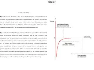
Figure 1-30
Figure 1-30. Cytotoxic CD8 T cells recognize antigen presented by class I MHC proteins. CD8 is a T cell coreceptor that binds to class I MHC molecules. CD8. TCR. class I MHC. Class II MHC proteins.
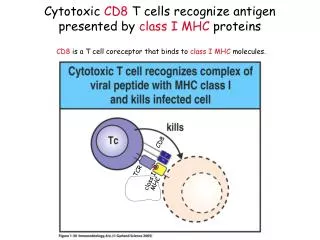

Figure 1-30
E N D
Presentation Transcript
Figure 1-30 Cytotoxic CD8 T cells recognize antigen presented by class I MHC proteins CD8 is a T cell coreceptor that binds to class I MHC molecules. CD8 TCR class I MHC
Peptides that bind to class II MHC molecules are longer than class I MHC-binding peptides
Figure 3-19 Expression of MHC class I versus class II proteins Class I MHC molecules are expressed on nearly all cells in your body.Class II MHC molecules are expressed on a subset of your cells -- mainly antigen presenting cells and B cells.Human class II MHC molecules are encoded by three loci: HLA-DR, HLA-DP, and HLA-DQ. Like class I MHC, there are hundreds of possible alleles in the human population for each type of class II MHC protein.
Figure 1-31 CD4 TH (T helper) cells activate macrophages or activate B cells to make antibody by recognizing antigen bound to class II MHC proteins CD4 is a T cell coreceptor that binds to class II MHC molecules. TH2 CD4 CD4 TCR TCR class II MHC class II MHC Levels of CD4 T cells are severely reduced in AIDS patients.
What does it mean that CD4 T cells “help” B cells make antibody and how are class II MHC molecules involved? Antigen (peptide) is presented by MHC class II on an APC to a CD4 TH cell with a TCR that recognizes a particular MHC class II/peptide complex. The TH cell is stimulated to undergo clonal expansion. If it encounters a B cell with the same class II/MHC peptide complex on its surface, it stimulates that B cell to clonally expand and produce soluble antibody.
Antigen presenting cells (APCs): can uptake any Ag because uptake is not receptor mediated. • B cells: mainly uptake only Ags that bind to B cell receptor (membrane Ig) -- result: TH only helps B cells that will make Ab against relevant Ag. • Explains hapten-carrier effect.
Clicker question In Douglas Adam’s famous novel Ultimate Answer to Life, the Universe, and Everything (a sequel to The Hitchhiker’s Guide to the Galaxy), a computer named Deep Thought is constructed in order to determine the ultimate answer to life, the universe, and everything. After seven and half million years of calculations, the computer answers: 1) “Quoting one is plagiarism. Quoting many is research.” 2) “All generalizations are false.” 3) “Common sense isn't.” “42.” “I have seen the truth and it makes no sense.” “43% of all statistics are useless.”
Clicker question In Douglas Adam’s famous novel Ultimate Answer to Life, the Universe, and Everything (a sequel to The Hitchhiker’s Guide to the Galaxy), a computer named Deep Thought is constructed in order to determine the ultimate answer to life, the universe, and everything. After seven and half million years of calculations, the computer answers: 1) “Quoting one is plagiarism. Quoting many is research.”* 2) “All generalizations are false.”* 3) “Common sense isn't.”* “42.” “I have seen the truth and it makes no sense.”* “43% of all statistics are useless.”* *http://www.deathstar.org/~fazzari/quotes.html
If you remember that the ultimate answer to life is “42”, you will remember that: CD4 T cells recognize class II MHC proteins.If you remember that you are 18 (or at least some of the freshman in Bi1 are 18), you will remember that: CD8 T cells recognize class I MHC proteins
Remember this: CD8 (T cell coreceptor and name for a class of T cells) Cytotoxic T lymphocyte (CTL) or killer T cell Class I MHC protein (HLA-A; HLA-B; HLA-C) Binds peptides derived from endogenous proteins CD4 (T cell coreceptor and name for a class of T cells)Helper T cell (TH) (further sub-divided into TH1 and TH2)Class II MHC protein (HLA-DR; HLA-DP; HLA-DQ) Binds peptides derived from exogenous proteins HIV infects CD4 T cells.
Figure 3-11 A T-cell receptor (TCR) looks like a membrane-bound version of an antibody Fab
TCR diversity • Junctional diversity -- combinatorial joining of gene segments, similar to antibody V,D,J • TCR a chains: V and J gene segments (like Ig light chains) • TCR b chains: V, D, and J gene segments (like Ig heavy chains) • Imprecise joining of V-J, V-D, and D-J • Combinatorial association of protein subunits (any a with any b) • NO SOMATIC MUTATION • Why?TH cells help B cells. B cell clones exist with anti-self antibody, but are not helped because TH cell is either eliminated or inactivated. If TCRs could undergo somatic mutation, they might mutate to have anti-self reactivity.
A6 TCR Ca Cb Va Vb CDR1 CDR2 CDR3 Garboczi et al., 1996, Nature 384:134; Ding et al., 1999, Immunity 11: 45. Animation by A.M. Giannetti
This is a TCR/class I MHC/peptide complex. TCRs bound to class II MHC/peptides look very similar. Ca,Cb A6 TCR CDR1 CDR2 CDR3 Va,Vb Tax peptide a1-a2 HLA-A2 Garboczi et al., 1996, Nature 384:134; Ding et al., 1999, Immunity 11: 45. b2m, a3 Animation by A.M. Giannetti
Clicker question 1) The bound peptide 2) Residues in the polymorphic top half of the protein 3) The non-polymorphic membrane-proximal domains Antibodies against foreign MHC molecules can be isolated from the blood of transplantation patients and multiparous women (women who have had more than one baby). Which portion of the MHC molecule are the antibodies likely to recognize? Polymorphic Less polymorphic or non-polymorphic
Clicker question Diversity results from combinatorial joining of gene segments and somatic mutation (not germline encoded): ___________ Diversity results from combinatorial joining of gene segments (not germline encoded): _____________ Diversity results from inheritance of different alleles (germline encoded): ______________ T cell receptors, MHC, antibodies MHC, antibodies, T cell receptors Antibodies, T cell receptors, MHC T cell receptors, antibodies, MHC MHC, T cell receptors, antibodies Antibodies, MHC, T cell receptors
T cell co-receptors • CD8 binds to MHC class I proteins. CD8 T cells are cytotoxic T lymphocytes (CTLs) or killer T cells. • CD4 binds to MHC class II proteins. CD4 T cells are “helper” T cells. • Two distinct functions of T cell co-receptors: Adhesion molecules - bind to appropriate MHC molecule (class I versus class II) on antigen presenting cell or infected cell and increase efficiency of antigen recognitionSignaling molecules - transduce signal as part of activation pathway
Clicker question CD4 and CD8 bind to class II and class I MHC molecules, respectively. Which part of an MHC/peptide complex are they likely to recognize? 1) The bound peptide 2) The a-helices on the peptide binding groove 3) The non-polymorphic membrane-proximal domains Polymorphic Less polymorphic or non-polymorphic
Unlike T cell receptors, CD4 and CD8 do NOT recognize peptides bound to MHC proteins Both co-receptors bind to a non-polymorphic region of an MHC protein distant from the peptide-binding site. CD4 binds to ALL class II MHC/peptide complexes. CD8 binds to ALL class I MHC/peptide complexes
A T cell receptor (TCR) and a T cell co-receptor bind simultaneously to the same MHC/peptide complex CD8b b2m Class I a1, a2, a3 CD8a TCR Va TCR Vb Model for TCR/classI/peptide/CD8 complex Target cell T cell From Gao et al., 1997, Nature 387, 630-634.
Model of CD4 and T cell receptor binding to a class II MHC/peptide complex Antigen presenting cell CD4 Class II MHC T cell receptor T cell Wang et al., 2001, PNAS 98, 10799-10804
CD4 also binds to HIV gp120, a component of the trimeric HIV envelope spike. This is a crystal structure of gp120 bound to CD4 and a Fab from an anti-gp120 antibody. All current crystal structures are of monomeric gp120. Kwong et al., 1998, Nature 393: 648-659
Clicker question • Which is true? • 1) Class II MHC CD8 T cells killer • 2) Class II MHC CD4 T cells helper • 3) Class I MHC CD4 T cells helper • 4) Class III MHC CD8 T cells cytotoxic • 5) Class III MHC CD2 T cells helpful
Large change in CDR3b upon binding to pMHC Reiser et al., 2002, Immunity 16: 345-354
T cell receptor complex • TCR ab chains are variable -- recognize MHC plus peptide • CD3 chains are invariable -- involved in signaling • TCR ab associates with CD3 in the membrane of a T cell
Cloning of TCR genes Cloning strategy based on the following assumptions: • TCRs should be expressed in T cells, but not in B cells • Messenger RNAs for TCRs will be found on membrane-bound polysomes • Genes encoding TCR should rearrange (like antibody genes) • TCR genes should have constant and variable regions.
Subtractive hybridization to clone TCR • Make library of T cell specific cDNAs (subtract out those cDNAs also expressed in B cells). • Make labeled cDNA as probe (made from membrane-bound poly(A)+ RNA). • Pull out T cell specific clones from library. • Look for rearrangement -- Southern blot genomic DNA from T cells versus cells in germline configuration (from non-immune cells).
Peptide binding to a class II MHC molecule Pockets for anchor residues marked by * Note both ends of groove are open. From Stern & Wiley (1994) Structure 2: 245-251
Peptide binding to a class I MHC molecule Pockets for anchor residues marked by * Note both ends of groove are closed. From Stern & Wiley (1994) Structure 2: 245-251
What does it mean for a T cell to “help” a B cell make antibody and how are class II MHC molecules involved? To understand the answer to this question, you need to know that: Class II MHC molecules on antigen presenting cells acquire peptides from exogenous* proteins. Exogenous proteins taken into antigen presenting cells end up in degradative compartments (endosomes, lysosomes) where they are cut up into peptides. Class II MHC molecules on B cells acquire peptides from exogenous proteins that bind to their B cell receptor (membrane bound antibody). These are taken into the cell by receptor-mediated endocytosis, and they end up in degradative compartments (endosomes, lysosomes) where they are cut up into peptides. Stimulating a B cell to make antibody takes three types of cells: B cells, T cells, Antigen presenting cells. *Exogenous: derived from outside of the cell. Opposite of “endogenous”. MHC class I molecules acquire peptides derived from endogenous proteins (e.g., viral proteins) produced inside a cell.
HEAVY LIGHT Potential diversity in TCRs versus Abs TCR diversity is heavily skewed towards junctional regions (CDR3). 1988, Nature 334, 395-402. # of junctional combinations affects the number of possible CDR3 sequences. # of variable region combinations affects the number of possible CDR1 and CDR2 sequences.
Model for TCR binding to MHC/peptide complex Top view of peptide binding site of a class I MHC molecule. MHC helices are yellow; peptide is pink. CDR3 regions (pink) are in the center, CDR1 and CDR2 (yellow and blue) are on the sides. The most variable part of a TCR is at the center of the combining site. The most variable part of an MHC/peptide complex is the peptide. CDR3 CDR3