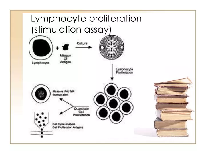
Lymphocyte proliferation (stimulation assay)
1.33k likes | 5.97k Views
Lymphocyte proliferation (stimulation assay). Direct cytotoxicity assay. PBMC. Mixed lymphocyte reaction. -Poor reproducibility - Radioactivity. Lymphocyte stimulation assay. Limiting dilution analysis. MLR Cytokine production, proliferation or cytotoxicity

Lymphocyte proliferation (stimulation assay)
E N D
Presentation Transcript
Direct cytotoxicity assay • PBMC
Mixed lymphocyte reaction -Poor reproducibility - Radioactivity
Limiting dilution analysis • MLR Cytokine production, proliferation or cytotoxicity - Aim: Yo predict alloreactive T cell precursors
Serology Based Typing –A panel of known anti-HLA antibodies are incubated with viable lymphocytes of unknown HLA type. –The HLA type of the sample is determined from the pattern of cell killing (cytotoxicity) that results from the antigen-antibody reactions. –Advantages •Rapid •Assesses HLA cell surface expression –Disadvantages •Limited detection of HLA polymorphism –low resolution •Requires viable cells •Requires HLA cell surface expression
Medium resolution HLA Typing (SSOP)SSOP: Sequence Specific Oligonucleotide Probe. –Genomic DNA is PCR amplified with primers that amplify all of the known alleles at each HLA locus (-A, -B, -C, DRB, -DQB). –PCR amplified sample DNA is hybridized with panels of probes directed against known polymorphisms. –HLA types are determined from the patterns of probe reactions. –SSOP provides partial sequence information. •Intermediate resolution. •Useful as a screening test. –Identifies family members that have inherited the same HLA chromosomes. –Identifies unrelated donors likely to be matched with the patient.–Also referred to as microarraytyping
High resolution HLA typing Sequence Based Typing (SBT) • Sample DNA is amplified by Polymerase Chain Reaction (PCR) • –Each HLA locus is amplified with either generic primers or allele specific primers. • Amplified DNA is used as a template for sequencing reactions using fluorescent dye-labeled dideoxynucleotides (ddATP, ddTTP, ddCTP, ddGTP). • Fluorescence labeled sequence fragments are analyzed by capillary gel electrophoresis, based on fragment size separation. • Sample sequence is compared with known HLA allele sequences to assign the HLA type. • –Method uses HLA sequencing analysis software. • –Method provides HLA allele level typing to determineallele matching and to detect rare or novel alleles.
Allo-Immune TestingDetection and characterization of recipient sensitization to HLAantigens. Patients with antibodies against a donor’s HLA antigens have an increased risk of rejection. -PRA(Panel Reactive Antibody) testing determines if the patient has antibodies against HLA antigens –Step 1: Screening test.»Patient serum is incubated with a panel of known HLA antigens todetermine if HLA antibodies are present in patient. –Step 2: Antibody Identification »Patient serum is incubated with individual HLA antigens to define the specificities of the patient’s HLA antibodies. –Crossmatch testing: Patient serum is incubated with donor cells. –Serology cross match. »Presence of donor specific antibodies in patient serum is determined from the pattern of cell killing (cytotoxicity) that results from the antigen-antibody reactions .–Flow Cytometry (FACS) crossmatch»This method further characterizes antibody type -IgGor IgM–against the donor
There are 14 KIR genes and two pseudogenes located in theleukocyte receptor complex (LRC) on chromosome 19q13.4. Human NK cells express various combinations of these 16 KIR genes with two common haplotypes: Group A, which has more inhibitory receptors and Group B, which has more activating receptors.
Standard STR/VNTR testingTests Informative loci (required before first standard assay) Indications To monitor engraftment afterallogeneic BMT* To detect relapse after allogeneic BMT* Maternal engraftment Genetic identity *Monozygotic twins excluded
Clinical Indications for Chimerism Testing in Hematopoietic Cell Transplant • Routine post-transplant documentation of the donor/recipient origin of white blood cells in peripheral blood and/or marrow. Documentation of engraftment may include testing lineage-specific cell subsets, such as CD3 positive T-cells and CD33 positive myeloid cells. • Evaluate donor/recipient cells in patients with inadequate marrow function. • Define whether recurrent or new malignancy has originated from recipient or donor cells. • Assess prognostic risks of rejection and recurrent malignancy. • Document the persistence of donor cells post-transplant in patients with recurrent disease or prior to donor lymphocyte infusion (DLI). • Evaluate whether graft rejection has occurred in recipients that are candidates for a second transplant. • Differentiate the origin of donor cells in recipients who have received a second transplant with a different donor or a transplant with double cord blood units. • Detect the presence of maternal derived cells in patients diagnosed with Severe Combined Immuno-Deficiency (SCID). • Verify genetic identity of putative identical twins.