Chapter 6: Skeletal System Chapter 4: Skeletal Tissue
570 likes | 817 Views
Chapter 6: Skeletal System Chapter 4: Skeletal Tissue . Pages 116-124; 150 &152 84; 89-91. Lesson Objectives. Skeletal Discuss the functions of the skeletal system. Classify bone in the basis of their shape and location. Describe parts of long bone.

Chapter 6: Skeletal System Chapter 4: Skeletal Tissue
E N D
Presentation Transcript
Chapter 6: Skeletal SystemChapter 4: Skeletal Tissue Pages 116-124; 150 &152 84; 89-91
Lesson Objectives • Skeletal • Discuss the functions of the skeletal system. • Classify bone in the basis of their shape and location. • Describe parts of long bone. • Describe the histological features of bone tissue. • Explain the steps involved in bone formation. • Describe the factors involved in bone growth and maintenance. • Compare the principal structural and functional differences between male and female skeletons. • Tissues • Explain how connective tissue is classified.
FUNCTIONS OF BONE AND THE SKELETAL SYSTEM • Support • Framework of body; supports soft tissue; provides points of attachment for skeletal muscles (most) • Protection • Of internal organs • Assisting in movement • How? Muscle to bone attachment; muscles contracts pulls on bone muscle + bone = movement • Mineral homeostasis • Minerals stored in bone tissue (Ca and P); released on demand when needed to other body parts via blood stream • Production of blood cells Red Bone Marrow; connective tissue
Red Bone Marrow • Within connective tissue • Red bone marrow through hemopoiesis makes: • Red blood cells • White blood cells • Platelets • Fragment of cytoplasm enclosed in a cell membrane; lacks a nucleus • Found in circulating blood • Plays role in hemostasis (stoppage of bleeding) • Red bone marrow consists of: • Developing red blood cells • Adipocytes (fat cells composed mainly of fat tissue adipose) • Fibroblasts (large, flat cells that secrete matrix of extracellular material of aerolar (collagen and elastic tissue) and dense connective tissue • Macrophages (white blood cells of immune system)
Location of Red Bone Marrow • Developing bones of fetus and some adult bones • Pelvis • Ribs • Sternum • Vertebrae • Skull • Ends of arm and thigh bones
FUNCTIONS OF BONE AND THE SKELETAL SYSTEM • Support • Protection • Assisting in movement • Mineral homeostasis • Production of blood cells • Triglyceride storage • In adipose tissue or yellow bone marrow
Yellow Bone Marrow • Yellow bone marrow consists of adipose tissue and some blood cells • Stores triglycerides in its adipose tissue • Potential energy reserve • Adults most red bone marrow has changed to yellow bone marrow • Not found in newborns
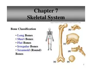
TYPES OF BONES • Four main types of bones of the body: • Long bones: have greater length and width, consists of a shaft and a variable number of ends, usually somewhat curved for strength. Examples: thigh (femur), leg (tibia and fibula), arm (humerus), forearm (ulna and radius), and fingers and toes (phalanges)
TYPES OF BONES • Four main types of bones of the body: • Short bones: somewhat cube-shaped and nearly equal in length and width. Examples: most wrist and ankle bones
TYPES OF BONES • Four main types of bones of the body: • Flat bones: generally thin, offer considerable protection, have extensive surface areas for muscle attachment. Examples: cranial bones (protect the brain), sternum/breastbone and ribs (protect organs in thorax), pelvis (protects digestive and reproductive organs),and scapulae (shoulder blades)
TYPES OF BONES • Four main types of bones of the body: • Irregular bones: complex shapes Examples: vertebrae of the backbone and some facial bones
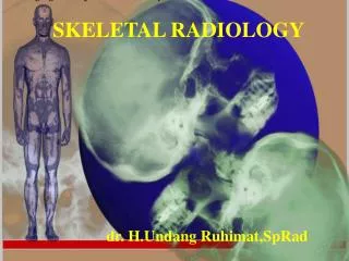
STRUCTURE OF BONE • Macroscopic Structure of Bone • Microscopic Structure of Bone
Macroscopic Structure of Bone • Parts as they relate to a long bone with greater length than width • Diaphysis: bone’s shaft or body; long, cylindrical, main portion of bone • Epiphyses: distal and proximal ends of bones • Metaphyses: regions in a mature bone where diaphysis joins epiphyses; in growing bone each metaphyses includes an epiphyseal plate, layer of hyaline cartilage that allows the diaphysis of bone to grow in length; when bone growth in length stops, cartilage in epiphyseal plate replaced by bone and this bony structure is now known as the epiphyseal line
Macroscopic Structure of Bone • Articular cartilage: thin layer of hyaline cartilage covering part of the epiphysis where bone forms a joint (articulation) with another bone; it reduces friction and absorbs shock; lacks a perichondrium so repair of damage is limited
Macroscopic Structure of Bone • Periosteum: tough sheath of dense irregular connective tissue surrounding a bone’s surface wherever it lacks articular cartilage; consists of bone-forming cells enabling bone to grow in diameter or thickness; protects the bone, assists in fracture repair, helps nourish bone tissue, serves as attachment point for ligaments and tendons
Macroscopic Structure of Bone • Medullary cavity: marrow cavity; space within diaphysis containing yellow bone marrow in adults • Endosteum: thin membrane lining medullary cavity; contains single layer of bone-forming cells
Microscopic Structure of Bone • Composition of intracellular materials of the bone, or osseous tissue matrix: • 25% water • 25% collagen fibers (protein) • 50% crystallized mineral salts • Calcification: mineral salts are deposited into a framework formed by collagen fibers they crystallize and tissue hardens • Calcification is initiated by: osteoblasts; bone-building cells_ • Hardness of bone depends on crystallized inorganic mineral salts • Flexibility depends on collagen fibers • Tensile strength is provided by collagen fibers and other organic molecules that offer resistance to being stretched or torn apart
Three Major Types of Bone Tissue • Osteoblasts=> blasts = buds or sprouts • Osteocytes => cytes = cells • Osteoclasts => clasts = break
Spaces between cells and matrix are for: • channels for blood vessels (supply bone with nutrients) • storage for red blood marrow • Composition of skeleton: BASED ON SIZE AND DISTRIBUTION OF SPACES 80% compact bone 20% spongy bone
Compact Bone Tissue • Contains few spaces; forms external layer of all bones; makes up bulk of diaphysis of long bone; provides protection and support; resists stress produced by weight and movement • Spongy Bone Tissue • Makes up most of the bone tissue of short, flat, and irregularly shaped bones; forms most of epiphyses of long bone and a narrow rim around the medullary cavity of the diaphysis of long bones.
Spongy Bone Tissue • Trabeculae: little beams; irregular latticework of thin columns of bone; spaces between filled with red bone marrow
Bone Scans • Darker spots = ‘hotspots’ • lighter spots = ‘coldspots’
OSSIFICATION: BONE FORMATION • Site of ossification: loose fibrous connective tissue membranes and pieces of hyaline cartilage, shaped like little bones in embryonic skeleton • First stage of ossification: appearance of osteogenic cells undergoing cell division to produce osteoblasts, which secrete matrix of bone • Ossification begins sixth/seventh week of embryonic life continues through adulthood
Two methods of bone formation • Intramembranous Ossification • directly on or within loose fibrous connective tissue membranes • Endochondral Ossification • within hyaline cartilage
Intramembranous Ossification Simplest of two methods -Forms flat bones of skull and mandible (lower jawbone) -Replaces ‘soft spot’ on fetal skull ______________
Endochondral Ossification • Replacement of hyaline cartilage by bone. • Most bone in the body below the skull except the clavicles are formed this way
HOMEOSTASIS OF BONE • Remodeling: ongoing replacement of old bone tissue by new bone tissue • osteoclasts responsible for the resorption(breaking down of bone matrix) • too much mineral deposited • surplus bone tissue may form thick bumps, called spurs • can interfere with joint movement • excessive loss of calcium • bone weakened • becomes overly flexible and vulnerable to fracture • Factors that control bone metabolism: • Minerals: Ca, P, Mg need adequate amounts of each • Vitamins: A, C, D • Hormones: hGH, IGFs (insulin-like growth factors; produced locally by bone and by the liver when stimulated by hGH ), insulin, thyroid hormones, parathyroid hormones, calcitonin • hGH: _main hormone before puberty that stimulates bone growth; produced by anterior lobe of pituitary gland • over secretion of hGH: produces giantism, person becomes taller and heavier than normal • under secretion of hGH: _produces dwarfism, short stature
Stress (weight bearing exercise): page 125 • Mineral crystals generate : production of collagen fibers, bone mass • Osteoblasts are: bone-forming cells • Heavily stressed bones are: notably thicker; builds & retains bone mass • Unstressed bones become: lose strength; loss of bone mass • Example: leg in cast up to ~30% of bone mass • Examples of serious bone health risks: • bedridden or paralyzed patients • people in weightless environments
Bone’s Role in Calcium Homeostasis • Bone stores 99% of the total amount of calcium in the body. • Calcium become available to other tissues when broken down during remodeling (replacement of old bone with new bone) • Effects of small changes in blood calcium levels: • Too high: heart may stop • Too low: breathing may cease
Bone’s Role in Calcium Homeostasis • Nerve cell functions depend on the right levels of Ca2+ • Enzymes require Ca2+, as a cofactor (non-protein component of enzymes bound to proteins and required for biological functions). • Blood clotting requires Ca2+. • Function of bone in calcium homeostasis in blood calcium levels • to “buffer” the blood calcium level, releasing Ca2+ to the blood when blood calcium levels falls and depositing Ca2+ back in bone when blood level rises • When levels falls in blood • parathyroid hormone (PTH) regulates Ca2+ and is produced by parathyroid glands • When levels rise in blood • calcitonin (CT) produced by thyroid gland
Negative Feedback System • Regulation of blood calcium (Ca2+) levels
Aging and the Skeletal System • Birth to adolescence=> more bone produced than lost • Young adults=> rate of production to loss about equal • Middle age=> decrease in bone mass • WHY??? • Levels of sex steroids lowered Greater problem for female; less bone mass to begin with = more osteoporosis in females Females age 30 bone loss starts; about age 45 estrogen levels decrease; by age 70 -30% of bone calcium lost Males begins around age 60, then loss of about 3% of bone mass every 10 years
COMMON DISORDERS---PAGE 152FRACTURES • fracture: any break in a bone • Four different types of fractures • (1) partial: incomplete break across the bone; i.e. a crack • (2) complete: complete break across the bone; bone broken in two or more pieces • (3) closed (simple): fractured bone does not break through skin • (4) open (compound): broken ends of bone protrude through skin
Fracture Repair What happens when a bone breaks? OUCH!!!
Blood Flow in Bones Note the blood vessels
Fracture Hematoma • Bleeding in the bone is stopped by a blood clot which closes off blood vessels • Lack of blood to bone kills osteocytes around fracture hematoma
Internal & External Callus • Previously inactive endosteum & periosteum undergo rapid mitosis & daughter cells migrate to area of fracture • Internal callus becomes spongy bone • External callus becomes cartilage 4. Bridge between bone fragments begins to form Internal callus
Osteoblasts replace cartilage w/spongy bone • Struts of spongy bone unite broken ends • Bone fragments are removed & replaced Bony callus of spongy bone
Remodeling • Can last from 4 months to over 1 year • May be unnoticeable or have a lump • Bone is usually thicker & stronger – a 2nd fracture seldom happens at the same site Healed fracture Obtain and read “Devil’s Pulpit Incident” and do the activity attached to it.
Connective Tissue Matrix • Matrix consists of fluid, gel, or solid ground substances plus protein fiber • Ground substance: component of connective tissue between cells and fibers, supports cells, binds them together, and provides a medium through which substances are exchanged between blood and cells • Fibers: strengthen and support connective tissues; three types embedded in matrix between cells: collagen fibers, elastic fibers, and reticular fibers
Three Types of Fibers • Loose connective tissue is composed of loosely woven collagen and elastic fibers. The fibers and other components of the connective tissue matrix are secreted by fibroblasts (i.e.: reticular fiber).
DUE: 11/18 EXTRA CREDIT: Sharks, whose skeletons are entirely made of cartilage, are being used in cancer research. Investigate the connection and write a small report. Be sure to include all references in MLA format….10 points