Download
1 / 28
280 likes | 309 Views
Explore the levels of structural organization, organs, and organ systems in the human body. Learn about homeostasis, control mechanisms, and feedback systems. Discover anatomical positions, body planes, and sections.

E N D
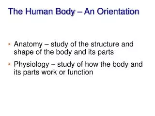
Overview of Anatomy and Physiology • Anatomy: Study of the structure/parts • Physiology: The study of function at many levels • Function always reflects structure; What a structure can do depends on its specific form
Levels of Structural Organization • Chemical: atoms and molecules • Cellular: cells and their organelles • Tissue: groups of similar cells • Organ: contains two or more types of tissues • Organ system: organs that work closely together • Organismal: all organ systems
Organelle Atoms Molecule Smooth muscle cell Cellular level 2 Chemical level 1 Smooth muscle tissue Cardiovascularsystem Tissue levelTissues consist of similartypes of cells. 3 Heart Bloodvessels Blood vessel (organ) Smooth muscle tissue Connective tissue Epithelialtissue Organ levelOrgans are made up of different typesof tissues. 4 Organismal levelThe human organism is made upof many organ systems. Organ system levelOrgan systems consist of differentorgans that work together closely. 6 5 Figure 1.1
Homeostasis • Definition: Maintenance of a relatively stable internal environment despite continuous outside changes • Homeostasis is maintained by homeostatic control mechanisms which involve at least three components: receptor, control center, effector
Components of a Homeostatic Control Mechanism • Receptor (sensor) • Monitors the environment and senses stimuli • Control center • Receives input from receptor • Determines the set point at which the variable is maintained • Determines appropriate response • Effector • Receives output from control center • Provides the means to respond • Response acts to reduce or enhance the stimulus (feedback)
4 Output:Information sent alongefferent pathway toeffector. 3 Input: Informationsent along afferentpathway to controlcenter. ControlCenter Afferentpathway Efferentpathway 2 Receptor Effector 5 Receptordetectschange. Responseof effectorfeeds backto reducethe effect ofstimulusand returnsvariable tohomeostaticlevel. 1 IMBALANCE Stimulusproduceschange invariable. BALANCE IMBALANCE Figure 1.4
Negative Feedback • When the response of a control mechanism reduces or shuts off/stops the original stimulus, this is called negative feedback • Example: • Regulation of body temperature
Control Center (thermoregulatory center in brain) Information sent along the afferent pathway to control center Information sent along the efferent pathway to effectors Efferent pathway Afferent pathway Receptors Temperature-sensitive cells in skin and brain Effectors Sweat glands Sweat glands activated Response Evaporation of sweat Body temperature falls; stimulus ends Stimulus Body temperature rises BALANCE Stimulus Body temperature falls Response Body temperature rises; stimulus ends Receptors Temperature-sensitive cells in skin and brain Effectors Skeletal muscles Afferent pathway Efferent pathway Shivering begins Information sent along the efferent pathway to effectors Information sent along the afferent pathway to control center Control Center (thermoregulatory center in brain) Figure 1.5
Positive Feedback • When the response of a control mechanism enhances or exaggerates the original stimulus, this is called positive feedback • Example: • Enhancement of labor contractions by oxytocin
Anatomical Position • Purpose: • Standard anatomical body position: • Body erect • Feet slightly apart • Palms facing forward
Upper limb Acromial Brachial (arm) Orbital Antecubital Nasal Antebrachial (forearm) Oral Carpal (wrist) Cervical Thoracic Axillary Digital Sternal Abdominal Lower limb Umbilical Coxal (hip) Pelvic Femoral (thigh) Inguinal Patellar Crural (leg) Fibular Pubic Tarsal (ankle) Thorax Abdomen Back (Dorsum) (a) Anterior/Ventral Figure 1.5
Upper limb Cephalic Acromial Occipital (back of head) Brachial (arm) Olecranal Cervical Back (dorsal) Scapular Vertebral Digital Lumbar Sacral Femoral (thigh) Gluteal Popliteal Sural (calf) Fibular Calcaneal Plantar (b) Posterior/Dorsal Figure 1.5
Body Planes and Sections • Sagittal plane • Divides body vertically into right and left parts • Produces a sagittal section • Midsagittal (median) plane • Lies on midline • Parasagittal plane • Not on midline
Body Planes • Frontal (coronal) plane • Divides body vertically into anterior and posterior parts • Transverse (horizontal) plane • Divides body horizontally into superior and inferior parts • Produces a cross section
Frontal plane Median (midsagittal) plane Transverse plane (a) Frontal section (through torso) (b) Transverse section (through torso, inferior view) (c) Median section (midsagittal) Pancreas Aorta Spleen Liver Spinal cord Intestines Rectum Spleen Left and right lungs Liver Heart Body wall Vertebral column Stomach Arm Subcutaneous fat layer Figure 1.6
Body Cavities • Two Large Cavities: • Dorsal cavity encloses the CNS • Two subdivisions: • Cranial cavity • Encases brain • Vertebral cavity • Encases spinal cord
Body Cavities • Ventral cavity • Houses soft internal organs (viscera) • Two subdivisions (separated by diaphragm): • Thoracic cavity • Abdominopelvic cavity
Cranial cavity Dorsal body cavity Ventral body cavity Cranial cavity Vertebral cavity Thoracic Cavity Dorsal body cavity Vertebral cavity Ventral body cavity (thoracic and abdominopelvic cavities) Diaphragm Abdominal cavity (contains digestive viscera) Abdomino- pelvic cavity Pelvic cavity (contains urinary bladder, reproductive organs, and rectum) (a) Lateral view (b) Anterior view Figure 1.7
Ventral Body Cavities • Thoracic cavity subdivisions: • Two pleural cavities • Each houses a lung • Mediastinum • Contains pericardial cavity • Also contains the esophagus and aorta • Pericardial cavity • Encloses heart
Ventral Body Cavities • Abdominopelvic cavity subdivisions: • Abdominal cavity • Contains stomach, intestines, spleen, and liver • Pelvic cavity • Contains urinary bladder, reproductive organs, and rectum
Cranial cavity Dorsal body cavity Ventral body cavity Cranial cavity Vertebral cavity Thoracic cavity Dorsal body cavity Vertebral cavity Ventral body cavity Diaphragm Abdominal cavity Abdomino- pelvic cavity Pelvic cavity (a) Lateral view (b) Anterior view Figure 1.7
Nine Abdominopelvic Regions Diaphragm Liver Right hypochondriac region Left hypochondriac region Epigastric region Stomach Gallbladder Transverse colon of large intestine Ascending colon of large intestine Right lumbar region Left lumbar region Umbilical region Descending colon of large intestine Small intestine Cecum Initial part of sigmoid colon Right iliac (inguinal) region Hypogastric (pubic) region Left iliac (inguinal) region Appendix Urinary bladder (a) Nine regions delineated by four planes (b) Anterior view of the nine regions showing the superficial organs Figure 1.12