Download
1 / 22
230 likes | 432 Views
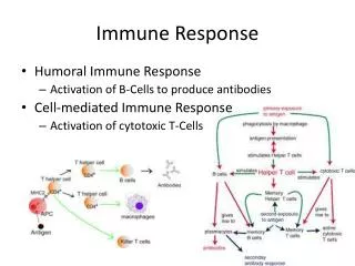
Role of Costimulation in Immune Response 2003. 4. 9. Yong-Hoon Chung, MD, PhD Department of Microbiology Hanyang Univ College of Medicine. Central Tolerance vs Peripheral Tolerance. B. Th. Y. FDC. APC. Costimulation of antigen and co-receptor activated T cells. SIGNAL 1:.

E N D
Role of Costimulation in Immune Response2003. 4. 9. Yong-Hoon Chung,MD, PhD Department of Microbiology Hanyang Univ College of Medicine
B Th Y FDC APC Costimulation of antigen and co-receptor activated T cells SIGNAL 1: Antigen Recognition Antigen-receptor and co-receptor ligation are often insufficient for proliferation and the expression of effector function
SIGNAL 2 Cognate T-professional APC co-stimulatory interaction APC activation induces B7 costimulatory molecules on B cells B7.1/2 APC Th Processing IL-1, IL-12 IFN- SIGNAL 1 Antigen recognition & co-receptor ligation Co-stimulation of T helper cells by professional APC CD28 Signal 1 & signal 2 are required for T cell clonal proliferation and differentiation to effector cells
High affinity IL-2 Receptor- and chains Low affinity IL-2 Receptor- and chains 1 1 2 IL-2 IL-2 IL-2 IL-2R IL-2R IL-2R Resting T cell Signal 1 Induction of high affinity IL-2R Signal 1 + 2 Increases IL-2 transcription and stabilises IL-2 mRNA Mechanism of co-stimulation in T cells
T cell Activated T cell - - - - Signal— 2 2 2 CD28 CD28 B7 B7 Cross-linking of CTLA-4 by B7 inhibits co-stimulation and inhibits T cell activation Costimulatory molecules also associate with inhibitory receptors Cross-linking of CD28 by B7 co-stimulates and induces CTLA-4 CTLA-4 binds CD28 with a higher affinity than B7 molecules to shut down the T cell response.
T T T CD28 B7 Professional APC presenting foreign antigen Epithelial cell expressing self antigen Epithelial cell expressing self antigen SIGNAL 1 AND 2 RESPONSE ANTIGEN SPECIFIC UNRESPONSIVENESS Co-stimulation in the induction of T cell anergy SIGNAL 1 ONLY
Fig. 1. PD-L¯PD-1 and other B7¯CD28 family members. The structures, interactions and functions of five of each of B7 ligand and CD28 family members are depicted schematically. aa indicates the number of amino acid residues of the cytoplasmic tail. Y represents a tyrosine residue.+ and - indicate costimulatory and coinhibitory signaling, respectively. Molecular weights are those of the glucosylated form. CD28, CTLA-4 & ICOS are homodimers. ind, inductive; M, monocyte; APC,antigen-presenting cell; EC, extracellular doman.
Fig. 2. Role of NKG2D engagement in human and mouse NK cells. (a) Ignorance: normal cells are spared from NK cell cytolytic activity, by signals (-) from MHC I specific inhibitory receptors(inh NKR, dark blue). Inhibitory NKR include CD158 (KIR-L) & CD85j(LIR1/ILT2) in humans, inhibitory Ly49 in mice & CD94¯NKG2A heterodimers in both species. In no stress, most cells don’t express ligands for NKG2D & same for ligands of NCR. Activating signals can be mediated, beside NCR, also by activating NKR; these are not shown but include activating CD158(KIR-S) in humans, Ly49D, Ly49H and Ly49P in mice and CD94¯NKG2C heterodimers in both species. (b) Tolerance: upon distress, cells may express only low levels of NKG2D ligands (NKG2D-L, light green), ligands of NCR (NCR-L, light red) & ligands for activating NKR(not shown). In these conditions, the inhibitory signal (-) initiated by engagement of inhibitory NKR overcomes activation signals (+). (c) Elimination: this can be mediated (i) via costimulation or (ii) after `primary recognition'. (i) Cellular distress inhibit MHC I expression & upregulation of NKG2D-L & also NCR-L(ligands of activating NKR). These modifications allow NK activation via NKG2D & NCR(NKR), which cooperate to overcome inhibitory signals mediated by NKR. (ii) Stressed cells expressing low levels of NCR-L (ligands of activating NKR), but hi levels of NKG2D-L, are eliminated by NK cells solely via NKG2D engagement.
Importance of ICOS–B7RP-1 costimulation in acute and chronic allograft rejectionÖzkaynak,E. et al.Nature Immunology2, 591 - 596 (2001) Figure 2.Blockade of ICOS–B7RP-1 costimulation inhibits acute allograft rejection. (a) The anti-ICOS mAb 12A8 did not deplete ICOS+ T cells, as shown by flow cytometric analysis of splenocytes 24 h after intraperitonal injection of mAb (200 g). Staining of splenocytes with a secondary (2°) antibody alone (mouse anti–rat IgG2b) detected anti-ICOS bound to CD3+ splenic T cells in treated, but not control, mice. Additional mAb increased T cell staining only marginally (upper right panel) and untreated mice had similar numbers of ICOS+ T cells (lower right panel) as animals receiving anti-ICOS therapy. (b) Northern analysis compared expression of ICOS and B7RP-1 RNA in control hearts versus cardiac allografts collected at day 7 after transplant from recipients treated with IgG or a blocking anti-ICOS (mAb 12A8). A murine GAPDH cDNA fragment was used as a control for loading (data are representative of three grafts per group). Beginning at the time of engraftment, wild-type (ICOS+/+) allograft recipients were treated daily for 14 days with (c) anti-ICOS or (d) ICOS-Ig fusion protein and monitored daily to determine the effects of therapy on allograft survival. Allograft survival in ICOS-/- recipients was also prolonged. Survival data are means.d. and are representative of six animals per group. *P<0.005 compared to other groups, as determined using Mann-Whitney U test
In vivo stimulation of CD137 broadens primary antiviral CD8+ T cell responses Halstead,E. S. et al.Nature Immunology3, 536 - 541 (2002) Figure 4.Effects of in vivo CD137 stimulation on the immunodominant NP(366–374) CD8+ T cell response in CD28-/- mice. Pulmonary lymphocytes were collected 10 days after intranasal infection with influenza virus and were stained with CD8 mAb and Db–NP(366–374) tetramers. (a) Representative flow cytometry plots are shown. Numbers indicate the percentages of CD8+ T cells that stained positive for the NP(366–374) tetramer. Pooled data from five independent experiments (n = 12 in each group) show cells that stained positive for CD8 and NP(366–374) tetramer as a measure of (b) total CD8+ T cells (c) total lymphocytes and (d) the absolute numbers of NP(366–374)-specific CD8+ T cells. (e) Representative experiment showing cytotoxicity of pulmonary lymphocytes against NP(366–374) peptide–loaded target cells.
Signaling through OX40 (CD134) breaks peripheral T-cell tolerancePratima Bansal-Pakala et al. Nature Medicine 7,907-912 (2001) Figure 2: OX40 signals, provided at the time of soluble antigen exposure, promote T-cell expansion and prevent induction of hypo-responsiveness.Recipients of V3/V11 CD4 T cells were tolerized with soluble peptide and treated with anti-OX40. a, Accumulation of V3/V11 CD4+ cells after 10 d in mice injected with PBS alone (bottom), or soluble peptide and either control IgG (middle) or anti-OX40 (top) given on day 2. Values represent means of 3 individual mice per group s.e.m. b, Accumulation of V3/V11 CD4+ cells after 17 d with mice challenged on day 10 with peptide in CFA. Mice initially injected with PBS (second bottom), or soluble peptide and either control IgG (second top) or anti-OX40 (top) given on day 2. Controls receiving PBS with no peptide/CFA challenge (bottom). Values represent means of 3 individual mice per group s.e.m. c and d, Proliferation (c) and IL-2 production (d) per V3/V11 CD4+ T cell after restimulation in vitro with peptide on day 17. Comparison between T cells from mice challenged on day 10, after initial injection with PBS alone () versus soluble peptide and either control IgG () or anti-OX40 (). IL-2 values are from pooled triplicate cultures. Similar results were seen in 2 separate experiments.
Inhibition of natural killer cells results in acceptance of cardiac allografts in CD28-/- mice Stefan Maier et al. Nature Medicine 7,557-562 (2001) Figure 5: Depletion of NK-receptor–bearing cells leads to prolonged survival of allogeneic cardiac grafts in CD28-/- recipients. a, Number of circulating DX5+CD3- NK cells in anti-NK1.1– or control-treated recipients. The percentage of NK cells was monitored in peripheral blood on day -4 (prior to antibody treatment) and on days 1 and 4 after treatment. b, Allograft survival in anti-NK1.1–treated H-2b, CD28-/- recipients (), anti-NK1.1–treated H-2b, CD28+/+ recipients () and control-treated H-2b, CD28-/- recipients () after transplantation of allogeneic (BALB/c, H-2d) cardiac grafts. c, Histological analysis of explanted allogeneic cardiac grafts after transplantation (as in Fig. 1b). Explantation of cardiac grafts was at day 5
Constitutive Early (activation) Late (activation) naïve T Cell CD28 LFA-1 CD2 CD40L CD27 OX40 4-1BB ICOS LIGHT APC ICAM-1 LFA-3 CD40 B7RP-1 B7/1 B7/2 CD70 CD40 OX40 L 4-1BB L ICOS L HVEM Cellular Expression of costimulatory molecules
Superfamily Positive Regulator NegativeRegulator Ig CD28 ICOS CD2 CTLA-4 PD-1 TNFR CD40 OX40 4-1BB LIGHT CD27 RANKL GITR Fas DR4/5 Cytokine TNFR IL-2 IL-15 IL-12 IL-1 IL-4 IL-10 Positive/NegativeRole of costimulatory molecules