RNA structure can be specific, stable and complex. (As a result, RNA mediates specific recognition and catalytic reac
Patterns and principles of RNA structure. RNA structure can be specific, stable and complex. (As a result, RNA mediates specific recognition and catalytic reactions.) Principles/ideas--RNAs contain characteristic 2° and 3° motifs Secondary structure--stems, bulges & loops


RNA structure can be specific, stable and complex. (As a result, RNA mediates specific recognition and catalytic reac
E N D
Presentation Transcript
Patterns and principles of RNA structure RNA structure can be specific, stable and complex. (As a result, RNA mediates specific recognition and catalytic reactions.) Principles/ideas--RNAs contain characteristic 2° and 3° motifs Secondary structure--stems, bulges & loops Coaxial stacking Metal ion binding Tertiary motifs (Pseudoknots, A-A platform, tetraloop/tetraloop receptor, A-minor motif, ribose zipper)
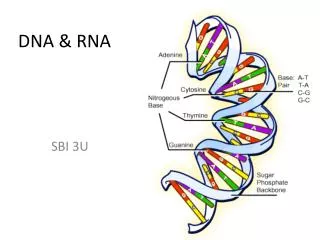
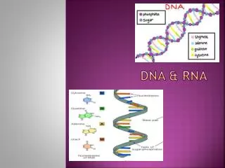
RNA vs. DNA nucleoside glycosidic bond nucleotide
RNA vs. DNA: who cares? Unstable backbone -OH Stable backbone Base-catalyzed RNA cleavage!
RNA transesterification mechanism transition state Base-catalyzed RNA cleavage! -OH + +
Different bases in RNA and DNA RNA only DNA only DNA and RNA
RNA chain is made single stranded! Chemical schematic One-letter code dsRNA can block protein synthesis and signal viral infections ssDNA can signal DNA damage and promote cell death Chain is directional. Convention: 5’ 3’.
Six backbone dihedral angles ()per nucleotide in RNA and DNA Is ssDNA floppy or rigid? Is RNA more or less flexible than ssDNA?
Two orientations of the bases: Anti and syn DNA and RNA Absent from undamaged dsDNA
-OH, what a difference an O makes! Different functions of DNA and RNA gene1gene2 gene3 . . . Stores genetic info Stores genetic info ssDNA signals cell death ssRNA OK E.g. mRNA = gene copy dsDNA OK dsRNA (“A” form) signals infection, mediates editing, RNA interference, . . . Double helical (B form) Forms complex structures Supercoiled Enzymes (e.g. ribosome), Binding sites & scaffolds Signals Templates (e.g. telomeres)
Examples of RNA structural motifs Stem, bulge, loop 4-helix junction Tetraloop Pseudoknot Sheared AA pairs Purine stacks Metal binding sites A-A platform Tetraloop receptor A-minor motif Ribose zipper . . . Secondary structures Tertiary structures
Cloverleaf representation of yeast Phe tRNA “Cloverleaf” conserved in all tRNAs Coaxial stacking of adjacent stems forms an L-shaped fold
Schematic drawing of yeast Phe tRNA fold Mg2+ (balls) Spermine
Non-WC base pairs and base triples in yeast tRNA Phe LOTS OF BASE COMBOS!! Enable alternate backbone orientations:
A9 intercalates between adjacent G45 and m7G46 in yeast tRNA Phe
Examples of RNA structural motifs Tetraloop Pseudoknot 4-helix junction Sheared AA pairs Purine stacks Metal binding sites A-A platform Tetraloop receptor A-minor motif . . .
UNCG tetraloop Stabilizes attached stem
HIV TAR RNA mediates Tat binding 2° structure schematic Coaxial stacking Base triple Nomenclature for secondary structure: stem, loop & bulge Arg binds GC bp
HIV TAR RNA mediates Tat binding 2° structure schematic Coaxial stacking Base triple Nomenclature for secondary structure: stem, loop & bulge
HIV TAR RNA mediates Tat binding 2° structure schematic Coaxial stacking Base triple Nomenclature for secondary structure: stem, loop & bulge Arg binds G26/C39 bp
Pseudoknots HDV ribozyme forms a double pseudoknot 1 2 1 Bases in loop of stem 1 form stem 2 (with bases outside stem 1)
Hepatitis Delta Virus (HDV) ribozyme double pseudoknot “Top” view 2° structure schematic U1A protein cocrystals
Hepatitis Delta Virus (HDV) ribozyme double pseudoknot “Top” view 2° structure schematic U1A protein cocrystals
Four-helix junction: L11 protein binding site in 23S RNA Four helices emerge from a central wheel. The four double-helical stems form two coaxial stacks. The two stacks have irregular but complementary shapes. The helices knit together to form a compact globular domain.
Base triples in the L11 4-helix junction • Bulge and loop mediate long-range tertiary interactions. • The riboses of A1084-A1086 (all A’s) form a “ribose zipper. • A1086 adopts a syn conformation to facilitate tight sugar packing.
Metal ions stabilize the L11 RNA 4-helix junction Mg2+ ions (gold balls) Cd2+ ions (magenta) Hg2+ (rose) RNA interactions of the central Cd2+ ion
P4-P6 Domain of the Group I ribozyme Two helical stacks are arranged parallel to each other. The structure is one helical radius thick. Two regions of 3° interactions between the two helical stacks. 1. Tetraloop/Tetraloop-receptor. 2. A-rich, single-stranded loop and the minor groove of the opposing helix.
Tertiary interactions in the P4-P6 domain Sheared AA Standard AU Sheared AA bps fill minor groove Cross-strand purine stack.
Tertiary interactions in the P4-P6 domain A-A platform Adjacent As pair side-by-side Side view Top view
Tertiary interactions in the P4-P6 domain A-A platform Adjacent As pair side-by-side Side view Top view
Metal ion core in the P4-P6 domain Divalent metal ions (Mg2+) are required for proper folding. These ions bind to specific sites and mediate the close approach of the phosphate backbones At one position in the molecule the phosphate backbone turns inward and coordinates two metal ions.
Adenosine-minor-groove base triples: the A-minor motif A fills minor groove & ribose 2’ OH forms H-bonds
Adjacent base-triples bring together RNA strands Hydrogen bonds between adjacent backbone atoms create a “ribose zipper” Deoxynucleotides destabilize P4-P6
The A-minor motif is widespread Conserved As are abundant in unpaired regions of structured RNAs. Group I intron P4-P6 % of As in “single-stranded regions
% of As in “single-stranded regions What happens in very large RNAs?
A-minor motifs are the predominant tertiary interaction in the 50S ribosomal subunit
Summary RNA structure can be specific, globular, stable and complex. (As a result, RNA mediates specific recognition and catalytic reactions.) 2. Secondary structures include stems, bulges, and loops. 3. Tertiary motifs include base triples, pseudoknots, A-A platforms, the tetraloop/tetraloop receptor, A-minor motifs, ribose zippers 4. Principles: stems and loops conserved, many non-WC base contacts, coaxial stacking, metal ion binding, H-bonding of ribose 2’ OH, and repeated “motifs”.