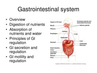
Gastrointestinal System Development
Gastrointestinal System Development. Suggested Reading: Langman’s 11 th ed. Ch. 14 (pp.209-231) --OR-- Langman’s 12 th ed. Ch. 15 (pp. 208-231). Matt Velkey matt.velkey@duke.edu 0380 Seely Mudd. Larsen’s fig 14-35. Gastrulation: Epiblast cells migrate through the

Gastrointestinal System Development
E N D
Presentation Transcript
Gastrointestinal System Development Suggested Reading: Langman’s 11th ed. Ch. 14 (pp.209-231) --OR-- Langman’s 12th ed. Ch. 15 (pp. 208-231) Matt Velkey matt.velkey@duke.edu 0380 SeelyMudd Larsen’s fig 14-35
Gastrulation: Epiblast cells migrate through the primitive streak. Definitive (embryonic) endoderm cells displace the hypoblast. Mesoderm spreads between endoderm and ectoderm. Langman’s fig 5.3
Cranial to caudal: Notochord (n) Paraxial mesoderm (pm) Intermediate mesoderm (im) *Lateral plate mesoderm (lpm) Extraembryonic mesoderm (eem) Early mesodermal patterning: (buccopharyngeal membrane) Specific regions of the epiblast migrate through the streak at different levels and assume different positions within the embryo: Langman’s fig 5-07
Endoderm Carlson fig 6-20 The developing endoderm (yellow) is initially open to the yolk sac (the cardiac region is initially most anterior)… Cranio-caudal folding at both ends of the embryo and lateral folding at the sides of the embryo bring the endoderm inside and form the gut tube.
Juxtaposition of ectoderm and endoderm at: Oropharyngeal (buccopharyngeal) membrane - future mouth Cloacal membrane - future anus Note: there actually isn’t much mesoderm in these membranes, which is important for their breakdown later in development to form the oral and anal orifices. cloacal membrane Carlson fig 6-20 Folding creates the anterior and posterior intestinal portals (foregut and hindgut, respectively) The cardiac region is brought to the ventral side of the developing gut tube.
Carlson fig 6-20 Gut-associated organs begin to form as buds from the endoderm: (e.g., thyroid, lung, liver, pancreas) Midgut opening to the yolk sac progressively narrows
Carlson fig 6-20 By the end of the first month: The stomach bulge is visible, Dorsal pancreas has begun to bud Connection of the midgut to the yolk sac is reduced to a yolk stalk and then a very thin vitelline duct
With lateral folding, mesoderm is recruited to gut wall Carlson fig 6-20 • Lateral folding of the embryo completes the gut tube • Mesodermal layer of the gut tube is called splanchnic (visceral) mesoderm - derived from lateral plate mesoderm • Somatic mesoderm lines body cavity Langman’s fig 6-18
Gut tube properDerivatives of gut tube Foregut: pharynx thyroid esophagus parathyroid glands stomach tympanic cavity proximal duodenum trachea, bronchi, lungs liver, gallbladder pancreas Midgut: proximal duodenum to right half of transverse colon Hindgut: left half of urinary bladder transverse colon to anus (These three regions are defined by their blood supply)
4th week 5th week Langman’s fig 14-4 Langman’s fig 14-14 Celiac artery supplies the foregut Superior mesenteric artery supplies the midgut Inferior mesenteric artery supplies the hindgut The figure on the right also shows the mesenteries; note also that the liver and stomach have dorsal and ventral mesenteries whereas the rest of the gut has only a dorsal mesentery.
A closer look at the mesenteries 5 weeks Last trimester • The stomach and liver are suspended in a mesentery that is attached to the dorsal AND ventral body walls • Dorsal mesentery of stomach becomes the greater omentum • Ventral mesentery of stomach/dorsal mesentery of the liver becomes the lesser omentum • Ventral mesentery of the liver becomes the falciform ligament • The rest of the GI tract is suspended in a dorsal mesentery (mesodoudenum, mesocolon, etc.) Langman’s fig 14-30 Langman’s fig 14-26
Regional patterning of the gut tube Gut = bilayered tube (endoderm surrounded by mesoderm) Regional gut tube patterning and organogenesis require bi-directional endoderm-mesoderm cross-talk and inductive signals from other nearby structures
Cranial-caudal pattern of the gut tube is played out as regional organ differentiation. Positional identity is assigned and distinct borders form. Esophageal/Gastric border Esophagus Stomach
Pyloric border (gastric/duodenal) Stomach Duodenum Duod. Stomach
Regional patterning of the gut tube - the Hox code Hox genes are evolutionarily conserved transcription factors that are used in regional patterning (flies to mammals). The gut has an cranial-caudal Hox gene expression pattern (code) similar to that seen in neural tissue. Some Hox genes are expressed in mesoderm, in overlapping patterns; some are expressed in endoderm. Hox gene expression boundaries correspond to morphologically recognizable elements in the GI tract. Hox gene expression is important for formation of major sphincters (red circles) Carlson fig 15-01
Hedgehog signaling is important for RADIAL (concentric) patterning of the entire gut tube Adult Esophagus Fetus Larsen’s fig 14-27 Wheater’s fig 14-5 • High hedgehog concentration directly inhibits smooth muscle differentiation (via repression of Smooth Muscle Activating Protein, or Smap) • Low Hedgehog concentration is permissive of muscle differentiation in the outer wall of the gut • High Hedgehog concentration also induces high BMP which inhibits neuron formation, thus limiting neurogenesis initially to the outer muscular wall of the gut (later in development, SHH goes away allowing development of the smooth muscle of the musularis mucosae and neurons of the submucosal plexus) Morphogen: induces different cell fates at different concentrations of signal
Regional Organogenesis: Esophagus Langman’s fig 14-06 • Region of foregut just caudal to lung bud develops into esophagus –errors in forming the esophagotracheal septa and/or re-canalization lead to tracheoesophageal fistulas and/or esophageal atresia, respectively. • Endodermal lining is stratified columnar and proliferates such that the lumen is obliterated; patency of the lumen established by re-canalization –errors in this process lead to esophageal stenosis • NOTE: this process of recanalization occurs throughout the gut tube, so occlusion can occur anywhere along the GI tract (e.g. duodenal stenosis) • Tube initially short and must grow in length to “keep up” with descent of heart and lungs –failure of growth in length leads to congenital hiatal hernia in which the cranial portion of the stomach is pulled into the hiatus. 7th week 5th week 9th week Larsen’s fig 14-22
Regional Organogenesis: Stomach 4th week • Stomach appears first as a fusiform dilation of the foregut endoderm which undergoes a 90° rotation driven by growth of the liver such that the left side moves ventrally and the right side moves dorsally (the vagus nerves follow this rotation which is how the left vagus becomes anterior and the right vagus becomes posterior). • Differential growth of the stomach borders establishes the greater and lesser curvatures; cranio-caudal rotation tips the pylorus superiorly. 3rd month Langman’s fig 14-08
Regional Organogenesis: Stomach 5th week 6th week (QuickTime version) Greater omentum 3rd month 3rd month • 90° rotation driven by growth of the liver such that the left side moves ventrally and the right side moves dorsally (the vagus nerves follow this rotation which is how the left vagus becomes anterior and the right vagus becomes posterior). • Dorsal AND ventral mesenteries of the stomach are retained to become the greater and lesser omenta, respectively • Caudal end of the stomach separated from the duodenum by formation of the pyloric sphincter (dependent on factors such as SOX-9, NKX-2.5, and BMP-4 signaling) –errors in this process lead to pyloric stenosis. (QuickTime version) Langman’s figs 14-11, 12
Pyloric Stenosis Rather common malformation: present in 0.5% - 0.1% of infants Characterized by very forceful (aka “projectile”), non-bilious vomiting ~1hr. after feeding (when pyloric emptying would occur). NOTE: the presence of bile would indicate POST-duodenal blockage of some sort. Hypertrophied sphincter can often be palpated as a spherical nodule; peristalsis of the sphincter seen/felt under the skin. Stenosis is due to overproliferation / hypertrophy of pyloric sphincter… NOT an error in re-canalization. More common in males than females, so most likely has a genetic basis which is as yet undetermined.
Regional Organogenesis: Liver & Pancreas Langman’s fig 14-19 • Liver and pancreas arise in the 4th week from foregut endoderm in response to signals from nearby mesoderm • Pancreas actually has ventral and dorsal components, each specified in a different manner
Cardiac mesoderm and septum transversum specifies liver and ventral pancreas (sort of…) Wnt ventral pancreas Wnt • ALL foregut endoderm has the potential to develop into either liver or pancreas but local signals repress these fates (mesodermal Wnts ↓ Liver; mesodermal Wnts + endodermal Shh ↓ Pancreas) • FGFs and BMPs from cardiac mesoderm and septum transversum inhibit Wnt signaling, but the endoderm still expresses Shh and thus develops into liver. • Meanwhile, endoderm just caudal to liver bud is out of reach from the FGFs and BMPs but still sees Wnts, so the tissue does NOT become liver. Unlike the rest of the endoderm, it does NOT express Shh and so develops into the ventral pancreas. Langman’s fig 14-22
The dorsal pancreas is specified by signaling from the notochord Signaling from the notochord represses Shh in caudal foregut endoderm, which permits dorsal pancreatic differentiation. Larsen’s fig 14-05
Rotation of the duodenum brings the ventral and dorsal pancreas together Larsen’s fig 14-09 Aberrations in this process may result in an annular pancreas, which can constrict the duodenum. Also, since the dorsal and ventral pancreas arise by different mechanisms, it’s possible that one or the other may be absent in the adult. Larsen’s fig 14-10
Rotation of the duodenum also causes it and the pancreas to become SECONDARILY retroperitoneal 5th week 3rd month Secondarily retroperitoneal = a structure that was originally in the body coelom but then got pushed into the body wall during development Langman’s fig 14-11
Intraperitoneal vs. retroperitoneal vs. secondarily retroperitoneal Larsen’s fig 14-16
Development of the midgut and colon Herniation and rotation: • Growth of the GI tract exceeds volume of abdominal cavity so the tube herniates through umbilicus • While herniated, gut undergoes a primary rotation (fig B) of 90° “counterclockwise” (when looking at the embryo); this corresponds with the rotation of the stomach, and positions the appendix on the left. The primary rotation also brings the leftvagus n. to the FRONT (hence the change in its name to ANTERIOR vagus n. • With the growth of the embryo, the abdominal cavity expands thus drawing the gut tube back within the abdominal cavity and causing an additional, secondary rotation (fig C) of 180° CCW (positioning the appendix on the RIGHT) • Once in the abdominal cavity, the colon continues to grow in length, pushing the appendix to its final position in the lower right quadrant. • Note the attachment of the vitelline duct to the gut at the region of the ileum. The duct normally regresses during development, but not always….
Defects associated with gut herniation and rotation: vitelline duct abnormalities Langman’s fig 14-32 Vitelline duct abnormalities of some sort occur in ~2% of all live births. Note that these aberrant structures are almost always found along the ileal portion of the GI tract.
Defects associated with gut herniation and rotation: oomphalocoele Langman’s fig 14-31
Defects associated with gut herniation and rotation: abnormal rotation Langman’s fig 14-33 Absent or incomplete secondary rotation Reversed secondary rotation (90 CCW primary rotation occurs as usual but followed by abnormal 180 CW rotation. Net rotation is 90° CW; viscera are in their normal location, but note that the duodenum is anterior to the transverse colon)
Defects associated with gut herniation and rotation: volvulus and ischemia/atresia fibrous septum Carlson fig 15-13 Langman fig 14-34 Volvulus (left figure) : fixation of a portion of the gut tube to the body wall; subsequent rotation twists and constricts the gut tube. Ischemia/atresia (right figure): blood supply to a portion of the mid- or hindgut may become compromised during rotation or herniation leading to ischemia and loss (A), fibrosis (B), septation (C), or narrowing (D) of that portion.
Development of the hindgut Langman’s fig 14-36 imperforate anus anal atresia anoperineal fistula rectovaginal fistula (QuickTime version) • Derivatives of the hindgut include everything caudal to the distal 1/3 of the transverse colon. • At distalmost portion (sigmoid colon and rectum), urorectal septum derived from mesoderm divides cloaca into the anorectal canal and urogenital canals; cloacal membrane breaks down to form anal opening 2 potential problems can arise: • “TOO MUCH” mesoderm – imperforate anus • “NOT ENOUGH” mesoderm - atresia (B), and/or fistulas (C – F) rectourethral fistula rectovesical fistula Carlson fig 15-18
Neural crest cells from vagal and sacral regions colonize the gut and give rise to the enteric neurons of the myenteric and submucosalplexes: Hirschsprung’s Disease: failure of vagal neural crest migration and/or survival, usually affecting distal hindgut (there are some sacral crest-derived cells there, but they cannot make up for loss of vagal crest-derived cells)
Hirschprung Disease (congenital megacolon) • Occurs in ~1:5000 births • Caused by failure of vagal neural crest cells to migrate into a portion of the colon • Aganglionic region tonically constricted (role of myenteric ganglia is largely INHIBITORY) • Signs and symptoms include: • constipation or small, infrequent bowel movements • bilious vomiting 3-4 hours after feeding (if totally obstructed) • distension of upstream colon (“megacolon”) • Surgically repaired by removing affected region