Download
1 / 1
10 likes | 115 Views
This study presents Spectral Self-Interference Fluorescence Microscopy (SSFM) in 4Pi mode as an innovative approach to achieve nanometer-level axial precision in biological imaging. SSFM enhances the visualization of fluorescent molecules in living cells, particularly targeting sub-cellular structures like lipid bilayer membranes and DNA strands. By utilizing interference patterns created from direct and reflected emissions, the technique yields a unique spectral signature essential for locating fluorophores. The work contributes significantly to the CenSSIS initiative, focusing on advancing multispectral imaging techniques for precise biological applications.

E N D
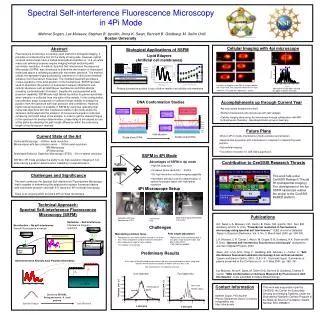
objective spacer mirror 16 14 12 10 5.1nm (white light) 8 3.4 nm (fluorescence) Height (nm) wl bottom 6 wl top 4 fl bottom 2 fl top Film of surface-absorbed water 0 2 nm 1 2 3 4 5 6 7 8 9 10 Position on the chip (mm) Silicon oxide spacer layer Silicon mirror Spectral Self-interference Fluorescence Microscopy in 4Pi Mode Mehmet Dogan, Lev Moiseev, Stephen B. Ippolito, Anna K. Swan, Bennett B. Goldberg, M. Selim Ünlü Boston University Cellular Imaging with 4pi microscope Abstract Biological Applications of SSFM Fluorescence microscopy is a widely used method in biological imaging. It provides a nondestructive tool for the study of living cells. However, optical confocal microscopes have a limited axial optical resolution of ~0.6 µm while many sub-cellular processes require imaging/vertical sectioning with nanometer resolution. A method, Spectral Self-interference Fluorescence Microscopy (SSFM), was introduced to determine the location of fluorescent molecules above a reflecting surface with nanometer precision. The method utilizes the spectral fringes produced by interference of direct and reflected emission from fluorescent molecules. The modified spectrum provides a unique signature of the axial position of the fluorophores. SSFM has been used to determine the position of fluorescent markers attached to sub-cellular structures such as lipid bilayer membranes and DNA strands revealing conformational information. Despite the unprecedented axial precision capability, SSFM lacks high lateral resolution for planar substrates when emission is collected from one side of the sample. In this configuration, low collection angle is required for sufficient fringe visibility to extract the position from the spectrum with high precision and confidence. However, higher lateral resolution is possible in SSFM by using two opposing high numerical objectives with two interference paths in 4pi mode where the sample is illuminated and the emission of fluorescence signal is collected coherently from both sides of the sample. In order to get the desired fringes in the spectrum for position determination, phase delay is introduced on one of the paths by adjusting the path length difference within the coherence length of the fluorescent markers. Lipid Bilayers (Artificial cell membranes) x-z slices of Shigella strain KAL O-antigen labeled with Texas Red. The separation between the slices is 483 nm. The scale bar represents 1 micron Probing fluorophore position in top or bottom leaflet of an artificial cell membrane Main lobe and side lobes marked for upper and lower cell walls Acomplishments up through Current Year DNA Conformation Studies • 4pi microscope designed and built. • Started characterizing the microscope with test samples • Cellular imaging done using 4pi microscope through collaboration with MPI for Biophysical Chemistry , Nanobiophotonics group in Germany average height of tags on hybridized DNA from SSFM average height of tags on single-strand from SSFM height of single-strand from white light height of double-strand from white light 12 nm 10 nm 8 nm 6 nm 12 nm 4 nm 10 nm 2 nm height of single-strand from white light 8 nm 6 nm Future Plans 4 nm 2 nm Hybridization Current State of the Art • Work in 4Pi-C mode: interference of both excitation and emission. • Spectral data acquisition with interference in collection for determining axial position . • Sub-cellular imaging. • Two-photon excitation for side lobe suppression. Double strand DNA Single strand DNA Confocal Microscopy : ~600nm axial resolution Microscopies with two collection arms: ~ 100nm axial resolution I5M Microscopy 4Pi Microscopy Stimulated Emission Depletion Microscopy (STED) :~30 nm lateral resolution SSFM in 4Pi mode provides the ability to do high resolution imaging in 3-D while having a position determination capability in axial dimension objective SSFM in 4Pi Mode Glass slide Advantages of SSFM in 4pi mode Contribution to CenSSIS Research Thrusts • High NA objectives • Increased lateral resolution : ~200nm • 3-D high resolution confocal imaging capability • Nanometer precision position determination of sparse fluorescent layers with high lateral resolution This work falls under CenSSIS Research Thrusts R1(multispectral imaging ). The development of the 4pi SSFM nanoscope relates the projret to the CenSSIS BioBED platform. Challenges and Significance This work combines the Spectral Self-interference Fluorescence Microscopy that is capable of determining the axial position sparse fluorescent layers with nanometer precision and high 3-D resolution 4Pi confocal microscopy. There is an ongoing effort to build a 4Pi confocal microscope 4Pi Microscope Setup Technical Approach: Spectral Self-interference Fluorescence Microscopy (SSFM) Publications A.K. Swan, L.A. Moiseev, C.R. Cantor, B. Davis, S.B. Ippolito, W.C. Karl, B.B. Goldberg, and M. S. Ünlü, "Towards nm resolution in fluorescence microscopy using spectral self interference,"IEEE Journal of Selected Topics in Quantum Electronics, Vol. 9, No. 2, March/April 2003, pp. 294-300. L.A. Moiseev, C.R. Cantor, I. Aksun, M. Dogan, B.B. Goldberg, A.K. Swan and M. S. Ünlü, “Spectral self-interference fluorescence microscopy” accepted to Journal of Applied Physics, 2004 Swan, A.K.; Unlu, M.S.; Tong, Y.; Goldberg, B.B.; Moiseev, L.; Cantor, C.; “Self-interference fluorescent emission microscopy 5-nm vertical resolution ”Lasers and Electro-Optics, 2001. CLEO '01. Technical Digest. Summaries of papers presented at the Conference on , 6-11 May 2001 ,pp. 360 -361 Lev Moiseev, Anna K. Swan, M. Selim Ünlü, Bennett B. Goldberg, Charles R. Cantor, “DNA Conformation on Surfaces Measured by Fluorescence Self-Interference”, to be submitted to Nature Biotechnology. Wide field imaging with Koehler Illumination Collection: APD and Spectrometer/CCD Interference Path Reflection – Self-interference Interference fringes in spectrum No reflection – No self-interference Smooth fluorescence envelope Challenges • Maintaining common focus • Two objectives must have common foci within 10-20 nm • Invar metal components used to minimize thermal drift • Ultra stable piezo stage for focus stability: • 1nm stability in closed loop • Path length adjustment • Right mirror in the interference path is • on a piezo stage for path length and • phase adjustments • λ/60 stepping capability Preliminary Results Self-interference Reveals Axial Position Information A thin layer of AlexaFluor488 on glass cover slip was scanned through the focus along axial direction where coherent excitation of 488nm CW laser was used (No interference in the collection channel) One Objective Two Objectives d2-d1=5nm Main Lobe FWHM=100nm Contact Information Mehmet Dogan, PhD Student Physics Department, Boston University mdogan@bu.edu http://ultra.bu.edu This work was supported in part by CenSSIS, the Center for Subsurface Sensing and Imaging Systems, under the Engineering Research Centers Program of the National Science Foundation (Award Number EEC-9986821) FWHM=660nm Best fit to MODEL fitting parameter: d, axial position Axial Position Spectral Fringes d2 d1