Download
1 / 42
470 likes | 777 Views
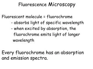
Fluorescence Microscopy Fluorescent molecule = fluorochrome - absorbs light of specific wavelength - when excited by absorption, the fluorochrome emits light of longer wavelength Every fluorochrome has an absorption and emission spectra. Fluorescence Microscopy.

E N D
Fluorescence Microscopy • Fluorescent molecule = fluorochrome • - absorbs light of specific wavelength • - when excited by absorption, the • fluorochrome emits light of longer • wavelength • Every fluorochrome has an absorption and emission spectra.
Fluorescence Microscopy Fluorochrome Structurally unstable when excited
EXCITATION SPECTRA Frequency of Event EMISSION SPECTRA Fluorochrome DAPI FITC Rhodamine Frequency of Event WAVELENGTH
Fluorescence Microscopy • Components: • Light source • Excitation filter • Emission filter • Dichroic mirror • -reflects short • -passes long SPECIMEN EYE PIECE
Frog Neuromuscular Junction Texas Red-Acetylated TUBULIN FITC ACTIN By Stephanie Moeckel-Cole
Flurochromes can be used in combination to mark different structures and/or molecules. “Multicolor "DiOlistic" labeling of the nervous system using lipophilic dye combinations.” Gan WB, Grutzendler J, Wong WT, Wong RO, Lichtman JW.
Labeled neurons in brain with different combination of fluorochromes. Fluorchromes: DiO DiI DiD
FLUORESCENCE MICROSCOPY • PROBLEM: Photobleaching (fading) • Photobeaching: Fluorochrome loses ability to fluoresce, absorb and emit light, due to damage or covalent modification. http://microscopyu.com/articles/fluorescence/fluorescenceintro.html
Fluorochrome PHOTOBLEACHING
(a-f) Images collected at 2 minute intervals. http://microscopyu.com/articles/fluorescence/fluorescenceintro.html
Quantum Dots : semiconductor nanoparticles, such as cadmium selenide, that emitted light after light excitation. • Advantages: brighter, no photobleaching, broad excitation • Disadvantages: potential toxicity for in vivo imaging Alivisatos et al.; Quantum dots as cellular probes.; Annu Rev Biomed Eng. 2005;7:55-76.
FLUORESCENCE MICROSCOPY • PROBLEM: Image degradation (blurring effect) due to light scattering http://www.microscopyu.com/articles/confocal/confocalintrobasics.html
CONFOCAL MICROSCOPY Light source: laser illumination with coherent light http://hyperphysics.phy-astr.gsu.edu/hbase/optmod/qualig.html#c4
CONFOCAL MICROSCOPY Collects light from one plane of the sample at a time Excludes out of focus light scatter
REGULAR FLUORESCENCE CONFOCAL MICRSCOPE MICROSCOPE
CONFOCAL MICROSCOPY Collect series of images from different focal planes Can assemble the image series to yield a 3-d image
Transmission Electron Microscopy (TEM) Very thin section Beam of electrons (=0.005 nm) Electromagnetic lenses Stain with metals Stain: electron dense: dark Unstained: light Nerve- osmium=myelin http://www.utsa.edu/tsi/assign/histo/Images/histst1.gif
Scanning Electron Microscopy (SEM) Surface structure Sectioning not required Metal coating of specimen Electron scattering Primary electrons Secondary electrons Detector http://www.chm.bris.ac.uk/pt/diamond/jamespthesis/chapter2_files/image002.gif
Ant Head Pollen
FREEZE FRACTURE Purpose: to analyze the distribution and density of integral membrane proteins in cell membranes Freeze a fragment of tissue Fracture using a sharp metal blade -fracture plane passes through lipid bilayers of a cell membrane Observe with SEM
FREEZE FRACTURE Cell Membrane: Lipid Bilayer EXTRACELLULAR SPACE CYTOPLASM FRACTURE EXTRACELLULAR SPACE CYTOPLASM
FREEZE FRACTURE EXTRACELLULAR SPACE Cell Membrane CYTOPLASM P face: inner face of inner membrane. EXTRACELLULAR SPACE CYTOPLASM E face: inner face of the outer membrane.
Intestinal Epithelium Microvilli Zonula adherens
Neuromuscular Junction • Synaptic site: • Active Zone: release • site of synaptic vesicles Heuser and Reese
STAINING • Histochemistry or Cytochemistry: dyes bind to certain types of molecules • Charged dyes bind to molecules of opposite charge
Charged dyes bind to molecules of opposite charge • Acidic Dye ---> dye- Eosin • Extracellular fibers, cytoplasmic filaments, and others • Basic Dye ---> dye+ ToluidineBlue • Alcian Blue • Cresyl Violet • Hematoxylin • Nuclei acids, glycosaminoglycans, ribosomes
Hematoxylin and Eosin Intestine
Staining Techniques • There are many dyes. • http://medinfo.ufl.edu/~dental/denhisto/stains.html • Examples: • Sudan black • -Lipids • Weigert Stain • -Reticular fibers Myelinated axons- blue ihcworld.com/imagegallery/displayimage.php?al...
Staining Techniques • Histochemical Stains: involve chemical reactions • Feulgen reaction • -DNA • Periodic Acid Shiff (PAS) • -neutral and acidic polysaccharides • - glycogen, mucous, basal laminae http://bioquant-com.bioquantusers.org/products.php?page=ls&content=gallery&sub=feulgen
Goblet cells PAS stain Intestinal Villus
Staining Techniques • Localization (staining) of an enzyme • AB + T AT + B ENZYME provide substrate • generate visible product
Staining Techniques • AB + T AT + B • Acetylcholinesterase- neuromuscular junction ACETYL CHOLINESTERASE Other stains for ATPases, alkaline phosphatases, and others
A technique to localize specific molecules in an organ, tissue or cell. IMMUNOCYTOCHEMISTRY
An organism creates antibodies to foreign molecules, ANTIGENS. An antigen may have different regions, EPITOPES, that are recognized as foreign by an organism. First, a bit of immunology……….
Polyclonal antibodies -A collection of distinct types of antibody molecules that recognize the same antigen (antibodies A + B + C) but often several different epitopes
Monoclonal antibodies -A single type of antibody molecule that recognizes only one epitope on an antigen (antibody A OR B OR C)