Two-Photon fluorescence Light Microscopy
550 likes | 1.33k Views
In The Name of GOD. An Introduction to:. Nano photonics Course Research. Dr S.M.Hamidi. Two-Photon fluorescence Light Microscopy. Mohammad Reza Sharifimehr. Laser and Plasma Research Institute-SBU. April 23,2013. 1/36. LAPRI-SBU. Two-Photon fluorescence Light Microscopy.

Two-Photon fluorescence Light Microscopy
E N D
Presentation Transcript
In The Name of GOD An Introduction to: Nanophotonics Course Research • Dr S.M.Hamidi Two-Photon fluorescence Light Microscopy Mohammad Reza Sharifimehr Laser and Plasma Research Institute-SBU April 23,2013. 1/36 LAPRI-SBU Two-Photon fluorescence Light Microscopy M.R.Sharifimehr
An Introduction to Microscopy Classes A brief history of TPA • Nature of Two-Photon Absorption (TPA) • Difference between TPA and SHG • Comparison of Two-Photon Absorption and One-Photon Absorption • The order of required intensity for Two-Photon Excitation (TPE) Outline • Design of a Two-Photon Microscope • Conventional Confocal and Two-Photon light microscopy • Advantages and Disadvantages of TPA • Recent TPA applications 2/36 LAPRI-SBU Two-Photon fluorescence Light Microscopy M.R.Sharifimehr
An Example of Microscopy • Scanning electron microscope (SEM) image of pollen 3/36 LAPRI-SBU Two-Photon fluorescence Light Microscopy M.R.Sharifimehr
Microscopy techniques has developed since 1590! Microscopy classes • 18th century microscopes 4/36 LAPRI-SBU Two-Photon fluorescence Light Microscopy M.R.Sharifimehr
Microscopes can be separated into several different classes. One grouping is based on what interacts with the sample to generate the image Optical (Photon) Microscopy (EM wave interaction with sample) Electron Microscopy (Electron wave interaction with sample) • Microscopy Scanning probe Microscopy Microscopy classes (force, voltage, current, … measurement, using a cantilever) Other types Common techniques 5/36 LAPRI-SBU Two-Photon fluorescence Light Microscopy M.R.Sharifimehr
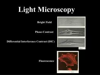
Optical (Photon) Microscopy (EM wave interaction with sample) White Light Microscopy Dark field microscopy Bright field microscopy Electron Microscopy (Electron wave interaction with sample) Polarized light microscopy Phase contrast microscopy Dispersion staining microscopy • Microscopy Scanning probe Microscopy Oblique illumination microscopy Microscopy classes (force, voltage, current, … measurement, using a cantilever) Other types Fluorescence Microscopy Confocal microscopy Two-photon excitation microscopy Light sheet fluorescence microscopy Common techniques 6/36 LAPRI-SBU Two-Photon fluorescence Light Microscopy M.R.Sharifimehr
Optical (Photon) Microscopy (EM wave interaction with sample) Electron Microscopy Transmission Electron Microscope (TEM) (Electron wave interaction with sample) Scanning Electron Microscope (SEM) Dark Field Microscopy • Microscopy Scanning probe Microscopy Microscopy classes (force, voltage, current, … measurement, using a cantilever) Other types Common techniques 7/36 LAPRI-SBU Two-Photon fluorescence Light Microscopy M.R.Sharifimehr
Optical (Photon) Microscopy (EM wave interaction with sample) Atomic Force Microscopy (AFM) Electrostatic Force Microscopy (EFM) Electron Microscopy Scanning Tunneling Microscopy (STM) (Electron wave interaction with sample) Scanning Thermal Microscopy (SThM) Scanning Capacitance Microscopy (SCM) • Microscopy Scanning probe Microscopy Scanning Near-Field Optical Microscopy (SNOM) Near-Field Scanning Optical Microscopy (NSOM) Microscopy classes (force, voltage, current, … measurement, using a cantilever) Other types Common techniques 8/36 LAPRI-SBU Two-Photon fluorescence Light Microscopy M.R.Sharifimehr
Optical (Photon) Microscopy (EM wave interaction with sample) Electron Microscopy (Electron wave interaction with sample) • Microscopy Scanning probe Microscopy Microscopy classes (force, voltage, current, … measurement, using a cantilever) Other types X-ray microscopy Scanning acoustic microscopes Ultrasonic Force Microscopy (UFM) Common techniques digital holographic microscopy (DHM) 9/36 LAPRI-SBU Two-Photon fluorescence Light Microscopy M.R.Sharifimehr
Optical (Photon) Microscopy (EM wave interaction with sample) Electron Microscopy (Electron wave interaction with sample) • Microscopy Scanning probe Microscopy Microscopy classes (force, voltage, current, … measurement, using a cantilever) Other types Microscope Image Processing Digital Microscopy (CCD+Microscope) Multifocal Plane Microscopy (Multiplane Microscopy) Common techniques 10/36 LAPRI-SBU Two-Photon fluorescence Light Microscopy M.R.Sharifimehr
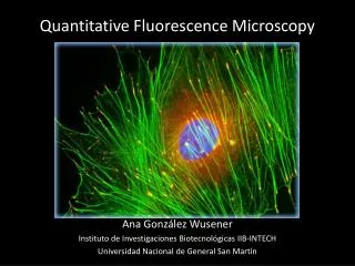
An Example of Fluorescence Microscopy • The microtubules are red • DNA is stained blue • A protein called INCENP is green 11/36 LAPRI-SBU Two-Photon fluorescence Light Microscopy M.R.Sharifimehr
+ + An Example of Fluorescence Microscopy fluorescent imaging of the human cancer cell 12/36 LAPRI-SBU Two-Photon fluorescence Light Microscopy M.R.Sharifimehr
Fluorescence Microscopy Schematic of a fluorescence microscope 13/36 LAPRI-SBU Two-Photon fluorescence Light Microscopy M.R.Sharifimehr
1929 Maria Goppert -Mayer- Theoretical basis of two-photon excitation was established 1963 Kaiser and Garret - two-photon excitation was verified experimentally 1990 Denk et al. - The invention of two-photon fluorescence light microscopy A brief history of Two-photon fluorescence light microscopy: Theory --> Experiment --> Application 14/36 LAPRI-SBU Two-Photon fluorescence Light Microscopy M.R.Sharifimehr
Milestones relevant to the development of two-photon microscopy A brief history of Two-photon fluorescence light microscopy: 15/36 LAPRI-SBU Two-Photon fluorescence Light Microscopy M.R.Sharifimehr
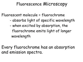
The familiar one-photon fluorescence process involves exciting a fluorophore from the electronic ground state to an excited state by a single photon. This process typically requires photons in the ultraviolet or blue/green spectral range. However, the same excitation process can be generated by the simultaneous absorption of two less energetic photons (typically in the infrared spectral range) under sufficiently intense laser illumination. This nonlinear processcan occur if the sum of the energies of the two photons is greater than the energy gap between the molecule’s ground and excited states. Under sufficiently intense excitation, three-photon and higher-photon excitation is also possible and deep UV microscopy based on these processes has been developed. Two-Photon Absorption and Fluorescence Definition 16/36 LAPRI-SBU Two-Photon fluorescence Light Microscopy M.R.Sharifimehr
Two Photon Absorption (TPA) Process Jablonski diagram of one-photon (a) and two-photon (b) excitation, which occurs as fluorophores are excited from the ground state to the first electronic states. One-photon excitation occurs through the absorption of a single photon. Two-photon excitation occurs through the absorption of two lower-energy photons via short-lived intermediate states. After either excitation process, the fluorophore relaxes to the lowest energy level of the first excited electronic states via vibrational processes. The subsequent fluorescence emission process for both relaxation modes is the same. 17/36 LAPRI-SBU Two-Photon fluorescence Light Microscopy M.R.Sharifimehr
Difference between TPA and SHG Two-Photon Excitation (TPE) is different from SHG, because in SHG the optical output is fixed at 2ω and is a very sharp line at 2ω, related to the line width only of the fundamental at frequency ω. In the TPE, ω3 is a higher frequency compared to either ω1 or ω2, but it is not 2ω1, 2ω2, or ω1 + ω2. In most cases, ω3 < (ω1+ ω2). 18/36 LAPRI-SBU Two-Photon fluorescence Light Microscopy M.R.Sharifimehr
TPA Equations 19/36 LAPRI-SBU Two-Photon fluorescence Light Microscopy M.R.Sharifimehr
One-photon and two-photon excitation are fundamentally different quantum-mechanical processes and have very different selection rules. A fluorophore that is one-photon active at wavelength λcan often be excited by two photons of twice the wavelength (2λ). A fluorophore’semission spectrum, is independent of the excitation mechanism, since the molecule relaxes to the same excited state through vibrational mechanisms before emission. Two (Multi)-Photon Absorption versus One-Photon Absorption A fluorophore’s two-photon excitation spectrum scaled to half the wavelength is typically not equivalent to its one-photon excitation spectrum. Further, fluorophores designed for one-photon excitation are not necessarily optimized for good two-photon absorption characteristics (such as σ). 20/36 LAPRI-SBU Two-Photon fluorescence Light Microscopy M.R.Sharifimehr
Two (Multi)-Photon Absorption versus One-Photon Absorption Comparison of one-photon (broken lines) and three-photon (solid lines) fluorescence excitation 21/36 LAPRI-SBU Two-Photon fluorescence Light Microscopy M.R.Sharifimehr
For a spatially uniform specimen, fluorescence signals are generated equally from each z-section above and below the focal plane for one-photon excitation. In contrast, over 80% of the total fluorescence signal can be confined to a region 1µm thick about the focal point using two-photon excitation. This property is a base for depth discrimination. Other differences between one-photon and two-photon excitation A demonstration of the localization of two-photon excitation volume 22/36 LAPRI-SBU Two-Photon fluorescence Light Microscopy M.R.Sharifimehr
Question: What’s the order of required intensity for two-photon excitation? Answer: A high-radiance light source on the order of is required for efficient two-photon excitation. High repetition rate (100 MHz), ultrafast (femtosecond or picosecond pulse widths) lasers, such as titanium–sapphire and Nd:YLF lasers, are the most widely used light sources. Two Photon Absorption (TPA) Process 23/36 LAPRI-SBU Two-Photon fluorescence Light Microscopy M.R.Sharifimehr
Conventional Light microscope --> Confocal Microscopy (1960s) --> Two-Photon Microscopy (1990s) --> 3D Imaging Confocal microscopy is a technique very similar to two-photon microscopy. The resolution of a microscope system scales inversely with the wavelength of light used. For a given fluorophore, two-photon excitation requires the use of excitation at twice the one-photon wavelength, resulting in approximately half the resolution. Confocal versus Two-Photon Light Microscopy Two-photon excitation wavelengths are typically about twice the one-photon excitation wavelengths. This wide separation between excitation and emission spectrum ensures that the excitation light and the Raman scattering can be rejected while filtering out a minimum of fluorescence photons. Near-infrared radiation used in two-photon excitation has orders of magnitude less absorption in biological specimens than UV or blue-green light. The attenuation of excitation light from scattering is also reduced, as the scattering cross-section decreases with increasing wavelength. 24/36 LAPRI-SBU Two-Photon fluorescence Light Microscopy M.R.Sharifimehr
Compared with confocal microscopy operating in the UV or blue-green excitation wavelengths, two-photon microscopy minimizes photobleaching and photodamage. The reduction in the photodamage volume results in a dramatic increase in viability of biological specimens. Confocal microscopy obtains 3D resolution by limiting the observation volume, whereas two-photon microscopy limits the excitation volume. Confocal versus Two-Photon Light Microscopy The reduction in the photodamage volume results in a dramatic increase in viability of biological specimens. 25/36 LAPRI-SBU Two-Photon fluorescence Light Microscopy M.R.Sharifimehr
? Barrier filter Galvo Mirrors Typical two-photon microscopy design A schematic drawing of typical components in a two-photon microscope. This system typically consists of a high-peak-power pulsed laser, a high-throughput scanning microscope and high-sensitivity detection circuitry. 26/36 LAPRI-SBU Two-Photon fluorescence Light Microscopy M.R.Sharifimehr
A critical component in a two-photon microscope is its light source and very kind of light sources can’t be use in this method. A High-sensitivity detection electronics, such as single-photon counting circuitry, is needed to ensure maximal detection efficiency. TPA Imaging Disadvantages Photodamage maybe caused by dielectric breakdown, resulting from the intense electromagnetic field of the femtosecond laser pulses. 27/36 LAPRI-SBU Two-Photon fluorescence Light Microscopy M.R.Sharifimehr
Concept--> Experiment --> Application My Experiment My Experiment 35/36 LAPRI-SBU Two-Photon fluorescence Light Microscopy M.R.Sharifimehr
3D Images are constructed by raster scanning the fluorescent volume in three dimensions using a galvanometer driven x–y scanner and a piezo-objective z-driver. A two-photon laser-scanning fluorescence microscopy 3D image of sulphorhodamine-stained human epidermis in vitro. A voxelgram generated from a two-photon image stack of a GFP-labeled cortical pyramidal neuron. Typical two-photon microscopy-Image Construction 28/36 LAPRI-SBU Two-Photon fluorescence Light Microscopy M.R.Sharifimehr
Carl Zeiss (over 160 years): Laser scanning microscope for multiphoton imaging TPA microscopes LSM 710 NLO & LSM 780 NLO http://microscopy.zeiss.com 29/36 LAPRI-SBU Two-Photon fluorescence Light Microscopy M.R.Sharifimehr
scanning microscope for multiphoton imaging Movie 1 TPA microscopes 29/36 LAPRI-SBU Two-Photon fluorescence Light Microscopy M.R.Sharifimehr
- Two-Photon Microscopy for 4D Imaging of Living Neurons - 3D Photonic crystals Fabrication using Two-Photon lithography - High-speed 3D printing Recent TPA Application - Single Molecule Detection 30/36 LAPRI-SBU Two-Photon fluorescence Light Microscopy M.R.Sharifimehr
Two-Photon Microscopy for 4D Imaging of Living Neurons Movies 2 Recent TPA Application 31/36 LAPRI-SBU Two-Photon fluorescence Light Microscopy M.R.Sharifimehr
3D Photonic crystals Fabrication using Two-Photon lithography Tightly focused in a photosensitive material, femtosecond laser pulses initiate two-photon polymerization and produce structures with a resolution down to 200 nm. Recent TPA Application SEM image of structures produced by two-photon polymerization of Deso-Bond 956-105. A resolution of ~200 nm was achieved. SEM image of a photonic crystal fabricated by two-photon lithography. From Cumpston et al. (1999) 31/36 LAPRI-SBU Two-Photon fluorescence Light Microscopy M.R.Sharifimehr
High-speed 3D printing of tiny objects: A race car model no larger than a grain of sand, created using the new high-speed two-photon lithography process Recent TPA Application A model of St. Stephen's Cathedral, Vienna, created using the new high-speed two-photon lithography process A model of London's Tower Bridge, created using the new high-speed two-photon lithography process 32/36 LAPRI-SBU Two-Photon fluorescence Light Microscopy M.R.Sharifimehr
Single Molecule Detection Single Molecules and Nanotechnology- R. Rigler-2008, Springer: Single emitting molecules are currently providing a new window into nanoscale systems ranging from biology to materials science. The amount of information that can be extracted from each single molecule depends upon the specific photophysical properties of the fluorophore and how these properties are affected by the nearby environment. Recent TPA Application Two-Photon Fluorescence scanning confocal image of single DCDHF-6 molecules in a PMMA film. 33/36 LAPRI-SBU Two-Photon fluorescence Light Microscopy M.R.Sharifimehr
Single-molecule spectroscopy has two requirements: 1-There is only one molecule present in the volume probed by the light source. This condition is met by using appropriate dilution together with microscopic techniques to probe a small volume. 2-The signal-to-noise (SNR) ratio for the single-molecule signal is sufficiently greater than unity for a reasonable averaging time to provide adequate sensitivity. For this purpose, large absorption cross sections, high photostability, operation below saturation of absorption, and (in the case of fluorescence detection) a high fluorescence quantum yield are needed. Recent TPA Application Fluorescence NSOM images of single molecules. 33/36 LAPRI-SBU Two-Photon fluorescence Light Microscopy M.R.Sharifimehr
We need a powerful software for Image processing Movies 3 References 34/36 LAPRI-SBU Two-Photon fluorescence Light Microscopy M.R.Sharifimehr
[1] Topics in Fluorescence Spectroscopy- Volume 5- Nonlinear and Two-Photon-Induced Fluorescence-Joseph R. Lakowicz-kluwer academic publishers [2] Two-Photon Fluorescence Light Microscopy-(2002),Peter TC So- ENCYCLOPEDIA OF LIFE SCIENCES / & 2002 Macmillan Publishers Ltd, Nature Publishing Group [3] Single Molecules and Nanotechnology- R. Rigler-2008, Springer [4] Multiphoton Fluorescence Light Microscopy -15th June 2012, KonstantinosPalikaras-John Wiley & Sons, Ltd [5] Nanophotonics- Paras N. Prasad, 2004, John Wiley & Sons- Chapters 3, 9, 11 [6] Properties of single organic molecules on crystal surfaces, Peter Grütter, 2006-Imperial College Press References [7] Nonlinear and Two-Photon-Induced Fluorescence, JOSEPH R. LAKOWICZ, 2002 Kluwer Academic Publishers [8] Photophysics of Molecular Materials, GuglielmoLanzani, 2006 WILEY-VCH Verlag GmbH [9] Photonics, Ralf Menzel, 2006 Springer [10] Nonlinear Optics, second edition, Robert W..Boyd, 2003 34/36 LAPRI-SBU Two-Photon fluorescence Light Microscopy M.R.Sharifimehr
There is a big difference between knowing and understanding. • For example, you can “know” the sky is blue but “understanding” why it’s blue is so much more important. Thanks 36/36 LAPRI-SBU Two-Photon fluorescence Light Microscopy M.R.Sharifimehr