Download
1 / 41
410 likes | 492 Views
Dive into the world of microscopy and learn about different types of microscopes, including electron and confocal laser scanning microscopes. Discover how these tools magnify objects and observe intricate details in various specimens. Explore the differences between viruses and cells.

E N D
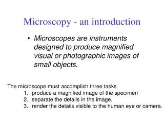
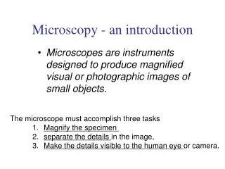
Microscopy - an introduction • Microscopes are instruments designed to produce magnified visual or photographic images of small objects. • The microscope must accomplish three tasks • 1. Magnify the specimen • separate the details in the image, • Make the details visible to the human eye or camera.
Microscopes increase resolution - minimumdistance two points can be apart and still be distinguished as two separate points
Microscope One or more lenses that make an enlarged image of an object.
Simple Microscope • Light passes through only 1 lens. • Example: magnifying glass
Compound Microscope • Lets light pass through an object and then through two or more lenses.
Stereoscopic Microscope • Gives a three dimensional view of an object. (Examples: insects and leaves) • Used for dissections
Electron microscopes – use a beam of electrons instead of a beam of light to magnify the image
Electron Microscopes • can achieve 3D images using electrons
The Scanning Electron Microscope • produces a 3-dimensional image of specimen’s surface features spider head of a butterfly
Scanning electron microscopy (SEM) • Types of specimens: • -Whole organisms • -Natural tissue surfaces • -Exposed tissue structure A flea magnified 50 000 X What is this?
Scanning Electron Microscope
Transmission electron microscopy (TEM). • Allows the observation of molecules within cells • Allows the magnification of objects in the order of 100, 000’s.
Longitudinal section of cilium Cross section of cilium 1 µm Transmissionelectronmicroscope (TEM) • Provides for detailed study of the internalstructure of cells • a beam of electrons is transmitted through the specimen for a 2D view Figure 6.4 (b) cilia on rabbit lungs
Transmission electron microscope Chloroplast from a tobacco leaf H1N1 virus
Confocal Laser Scanning Microscope (CLSM) • laser beam used to illuminate spots on specimen • computer compiles images created from each point to generate a 3-D image • used on specimens that are too thick for a light microscope
A, B, C pollen grains: Scanning electron microscope D pollen grains: Confocal Laser Scanning Microscope E pollen grains: Transmission electron microscope F pollen grains: Light microscope G Mixed pollen grains (bright field light microscope, stained) H pollen grains confocal laser scanning microscope
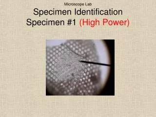
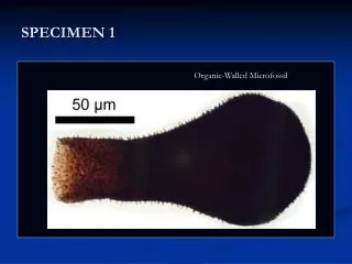
Look at the following micrographs (a picture made by a microscope) and try to determine what the object is!
What is the difference between a… VIRUS and CELL? E.coli bacterial cells
VIRUS BACTERIA • can’t live on its own- must - can exist on its own live inside another cell • much smaller (20 – 400nm) - larger (1000 nm = 1μm) • none are beneficial - some can be beneficial (bacteria in gut) • no cell wall, only a protein - outer cell wall coat - cannot be killed by antibiotics - are killed by antibiotics